8FUC

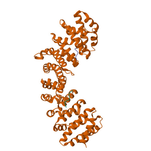
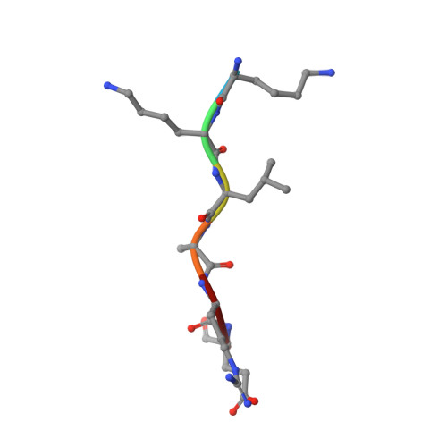
Crystal structure of mouse Importin alpha in complex with Hendra virus matrix protein minor site NLS2


Experimental Data Snapshot

  • Method: X-RAY DIFFRACTION
  • Resolution: 2.10 Å
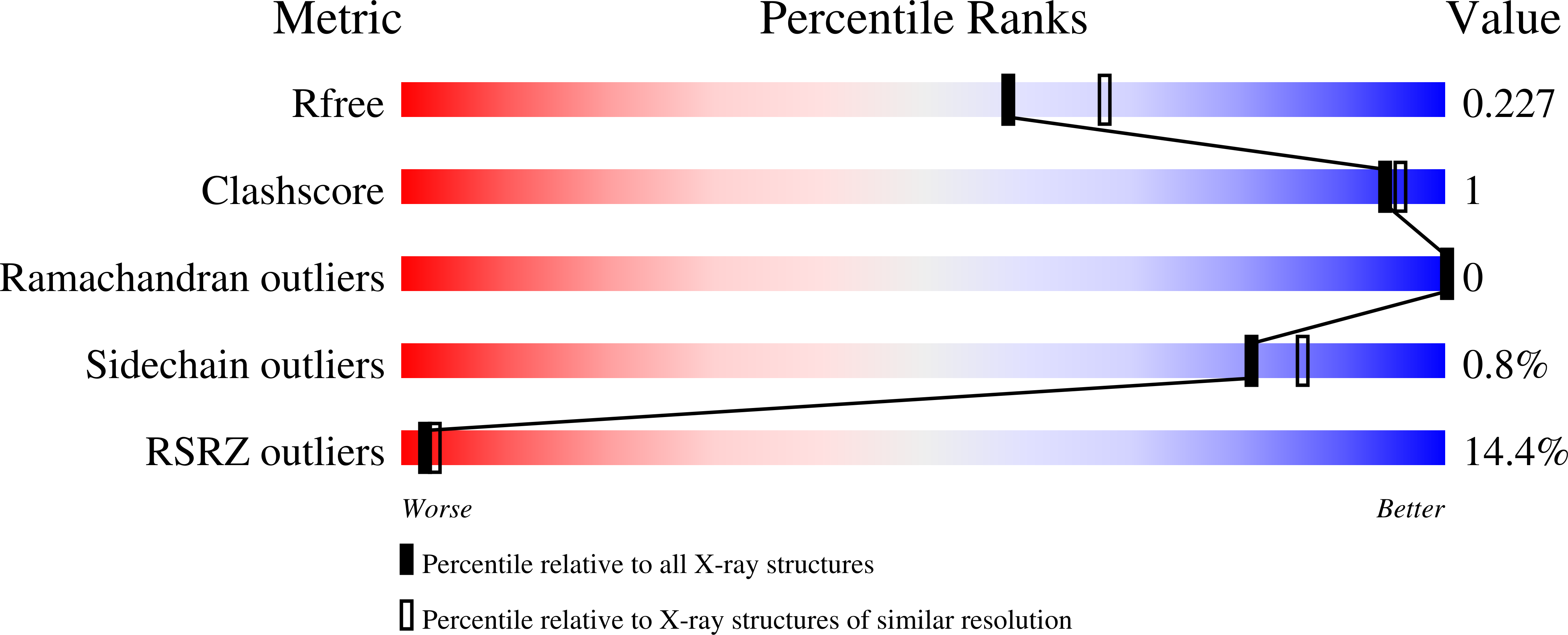
  • R-Value Free: 0.217 
  • R-Value Work: 0.175 
  • R-Value Observed: 0.177 

Starting Model: experimental
View more details

wwPDB Validation   3D Report Full Report


This is version 1.1 of the entry. See complete history


Literature

Henipavirus Matrix Protein Employs a Non-Classical Nuclear Localization Signal Binding Mechanism.

Donnelly, C.M.Vogel, O.A.Edwards, M.R.Taylor, P.E.Roby, J.A.Forwood, J.K.Basler, C.F.

(2023) Viruses 15

  • DOI: https://doi.org/10.3390/v15061302
  • Primary Citation of Related Structures:  
    8FUA, 8FUB, 8FUC

  • PubMed Abstract: 

    Nipah virus (NiV) and Hendra virus (HeV) are highly pathogenic species from the Henipavirus genus within the paramyxovirus family and are harbored by Pteropus Flying Fox species. Henipaviruses cause severe respiratory disease, neural symptoms, and encephalitis in various animals and humans, with human mortality rates exceeding 70% in some NiV outbreaks. The henipavirus matrix protein (M), which drives viral assembly and budding of the virion, also performs non-structural functions as a type I interferon antagonist. Interestingly, M also undergoes nuclear trafficking that mediates critical monoubiquitination for downstream cell sorting, membrane association, and budding processes. Based on the NiV and HeV M X-ray crystal structures and cell-based assays, M possesses a putative monopartite nuclear localization signal (NLS) (residues 82 KRKKIR 87 ; NLS1 HeV), positioned on an exposed flexible loop and typical of how many NLSs bind importin alpha (IMPα), and a putative bipartite NLS ( 244 RR-10X-KRK 258 ; NLS2 HeV), positioned within an α-helix that is far less typical. Here, we employed X-ray crystallography to determine the binding interface of these M NLSs and IMPα. The interaction of both NLS peptides with IMPα was established, with NLS1 binding the IMPα major binding site, and NLS2 binding as a non-classical NLS to the minor site. Co-immunoprecipitation (co-IP) and immunofluorescence assays (IFA) confirm the critical role of NLS2, and specifically K258. Additionally, localization studies demonstrated a supportive role for NLS1 in M nuclear localization. These studies provide additional insight into the critical mechanisms of M nucleocytoplasmic transport, the study of which can provide a greater understanding of viral pathogenesis and uncover a potential target for novel therapeutics for henipaviral diseases.


  • Organizational Affiliation

    School of Dentistry and Medical Sciences, Charles Sturt University, Wagga Wagga, NSW 2678, Australia.


Macromolecules

Find similar proteins by:  Sequence   |   3D Structure  

Entity ID: 1
MoleculeChains Sequence LengthOrganismDetailsImage
Matrix protein18Hendra virus horse/Australia/Hendra/1994Mutation(s): 0 
UniProt
Find proteins for O89341 (Hendra virus (isolate Horse/Autralia/Hendra/1994))
Explore O89341 
Go to UniProtKB:  O89341
Entity Groups  
Sequence Clusters30% Identity50% Identity70% Identity90% Identity95% Identity100% Identity
UniProt GroupO89341
Sequence Annotations
Expand
  • Reference Sequence
Find similar proteins by:  (by identity cutoff)  |  3D Structure
Entity ID: 2
MoleculeChains Sequence LengthOrganismDetailsImage
Importin subunit alpha-1510Mus musculusMutation(s): 0 
Gene Names: Kpna2Rch1
UniProt & NIH Common Fund Data Resources
Find proteins for P52293 (Mus musculus)
Explore P52293 
Go to UniProtKB:  P52293
IMPC:  MGI:103561
Entity Groups  
Sequence Clusters30% Identity50% Identity70% Identity90% Identity95% Identity100% Identity
UniProt GroupP52293
Sequence Annotations
Expand
  • Reference Sequence

Find similar proteins by:  Sequence   |   3D Structure  

Entity ID: 3
MoleculeChains Sequence LengthOrganismDetailsImage
Contaminant peptide KKLAREC [auth F]6Escherichia coli BL21(DE3)Mutation(s): 0 
Sequence Annotations
Expand
  • Reference Sequence
Experimental Data & Validation

Experimental Data

  • Method: X-RAY DIFFRACTION
  • Resolution: 2.10 Å
  • R-Value Free: 0.217 
  • R-Value Work: 0.175 
  • R-Value Observed: 0.177 
  • Space Group: P 21 21 21
Unit Cell:
Length ( Å )Angle ( ˚ )
a = 78.55α = 90
b = 90.18β = 90
c = 98.04γ = 90
Software Package:
Software NamePurpose
MOSFLMdata reduction
Aimlessdata scaling
PHASERphasing
REFMACrefinement
Cootmodel building

Structure Validation

View Full Validation Report


Entry History & Funding Information

Deposition Data


Funding OrganizationLocationGrant Number
National Institutes of Health/National Institute Of Allergy and Infectious Diseases (NIH/NIAID)United States5R21AI144880-02

Revision History  (Full details and data files)

  • Version 1.0: 2023-01-25
    Type: Initial release
  • Version 1.1: 2023-08-30
    Changes: Data collection, Database references