8TWT

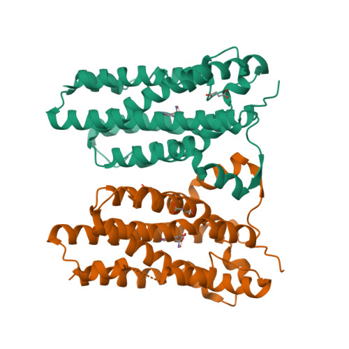
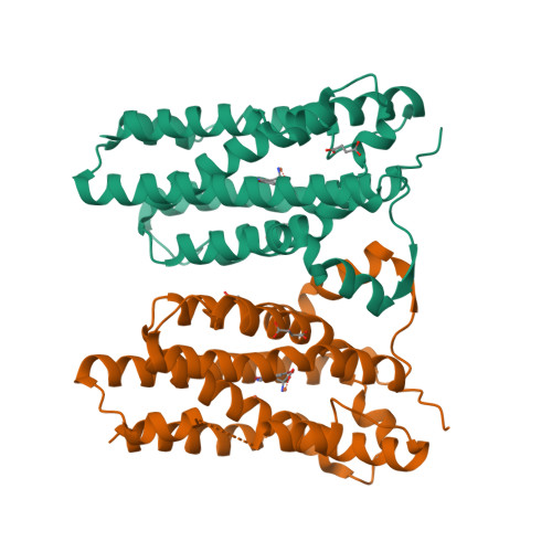
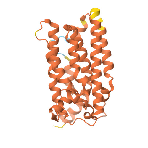
Crystal structure of nitrile synthase AetD with substrate bound and cofactor partially assembled

  • Classification: OXIDOREDUCTASE
  • Organism(s): Aetokthonos hydrillicola
  • Expression System: Escherichia coli
  • Mutation(s): No 

  • Deposited: 2023-08-21 Released: 2024-08-28 
  • Deposition Author(s): Ye, N., Drennan, C.L.
  • Funding Organization(s): Howard Hughes Medical Institute (HHMI), National Institutes of Health/National Institute of General Medical Sciences (NIH/NIGMS)

Experimental Data Snapshot

  • Method: X-RAY DIFFRACTION
  • Resolution: 2.30 Å
  • R-Value Free: 0.217 
  • R-Value Work: 0.184 
  • R-Value Observed: 0.186 

wwPDB Validation   3D Report Full Report


Ligand Structure Quality Assessment 


This is version 1.3 of the entry. See complete history


Literature

A single diiron enzyme catalyses the oxidative rearrangement of tryptophan to indole nitrile.

Adak, S.Ye, N.Calderone, L.A.Duan, M.Lubeck, W.Schafer, R.J.B.Lukowski, A.L.Houk, K.N.Pandelia, M.E.Drennan, C.L.Moore, B.S.

(2024) Nat Chem 16: 1989-1998

  • DOI: https://doi.org/10.1038/s41557-024-01603-z
  • Primary Citation of Related Structures:  
    8TWN, 8TWT, 8TWW

  • PubMed Abstract: 

    Nitriles are uncommon in nature and are typically constructed from oximes through the oxidative decarboxylation of amino acid substrates or from the derivatization of carboxylic acids. Here we report a third nitrile biosynthesis strategy featuring the cyanobacterial nitrile synthase AetD. During the biosynthesis of the eagle-killing neurotoxin, aetokthonotoxin, AetD transforms the 2-aminopropionate portion of 5,7-dibromo-L-tryptophan to a nitrile. Employing a combination of structural, biochemical and biophysical techniques, we characterized AetD as a non-haem diiron enzyme that belongs to the emerging haem-oxygenase-like dimetal oxidase superfamily. High-resolution crystal structures of AetD together with the identification of catalytically relevant products provide mechanistic insights into how AetD affords this unique transformation, which we propose proceeds via an aziridine intermediate. Our work presents a unique template for nitrile biogenesis and portrays a substrate binding and metallocofactor assembly mechanism that may be shared among other haem-oxygenase-like dimetal oxidase enzymes.


  • Organizational Affiliation

    Center for Marine Biotechnology and Biomedicine, Scripps Institution of Oceanography, University of California San Diego, La Jolla, CA, USA.


Macromolecules
Find similar proteins by:  (by identity cutoff)  |  3D Structure
Entity ID: 1
MoleculeChains Sequence LengthOrganismDetailsImage
AetD
A, B
260Aetokthonos hydrillicolaMutation(s): 0 
Gene Names: aetD
UniProt
Find proteins for A0A861B387 (Aetokthonos hydrillicola Thurmond2011)
Explore A0A861B387 
Go to UniProtKB:  A0A861B387
Entity Groups  
Sequence Clusters30% Identity50% Identity70% Identity90% Identity95% Identity100% Identity
UniProt GroupA0A861B387
Sequence Annotations
Expand
  • Reference Sequence
Small Molecules
Ligands 5 Unique
IDChains Name / Formula / InChI Key2D Diagram3D Interactions
67I (Subject of Investigation/LOI)
Query on 67I

Download Ideal Coordinates CCD File 
C [auth A],
G [auth B]
(2S)-2-azanyl-3-[5,7-bis(bromanyl)-1H-indol-3-yl]propanoic acid
C11 H10 Br2 N2 O2
UURVQGNSCUCIQS-VIFPVBQESA-N
MLT
Query on MLT

Download Ideal Coordinates CCD File 
D [auth A],
H [auth B]
D-MALATE
C4 H6 O5
BJEPYKJPYRNKOW-UWTATZPHSA-N
SIN
Query on SIN

Download Ideal Coordinates CCD File 
E [auth A]SUCCINIC ACID
C4 H6 O4
KDYFGRWQOYBRFD-UHFFFAOYSA-N
GOL
Query on GOL

Download Ideal Coordinates CCD File 
I [auth B]GLYCEROL
C3 H8 O3
PEDCQBHIVMGVHV-UHFFFAOYSA-N
FE
Query on FE

Download Ideal Coordinates CCD File 
F [auth A],
J [auth B],
K [auth B]
FE (III) ION
Fe
VTLYFUHAOXGGBS-UHFFFAOYSA-N
Experimental Data & Validation

Experimental Data

  • Method: X-RAY DIFFRACTION
  • Resolution: 2.30 Å
  • R-Value Free: 0.217 
  • R-Value Work: 0.184 
  • R-Value Observed: 0.186 
  • Space Group: P 21 21 21
Unit Cell:
Length ( Å )Angle ( ˚ )
a = 65.916α = 90
b = 86.089β = 90
c = 101.779γ = 90
Software Package:
Software NamePurpose
XDSdata reduction
XSCALEdata scaling
PHASERphasing
Cootmodel building
PHENIXrefinement

Structure Validation

View Full Validation Report


Ligand Structure Quality Assessment 


Entry History & Funding Information

Deposition Data


Funding OrganizationLocationGrant Number
Howard Hughes Medical Institute (HHMI)United States--
National Institutes of Health/National Institute of General Medical Sciences (NIH/NIGMS)United StatesR35-GM126982
National Institutes of Health/National Institute of General Medical Sciences (NIH/NIGMS)United StatesP30-GM124165
National Institutes of Health/National Institute of General Medical Sciences (NIH/NIGMS)United StatesP30-GM133894

Revision History  (Full details and data files)

  • Version 1.0: 2024-08-28
    Type: Initial release
  • Version 1.1: 2024-09-25
    Changes: Database references
  • Version 1.2: 2024-10-02
    Changes: Database references
  • Version 1.3: 2024-12-18
    Changes: Database references, Structure summary