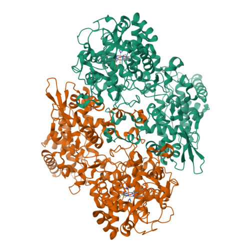
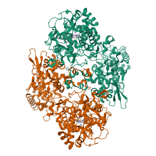
8W1X

2.35-angstrom resolution intermediate crystal structure of KatG from Mycobacterium tuberculosis with an MYW-OOH cofactor soaked with peroxide for 5 minutes


Experimental Data Snapshot

  • Method: X-RAY DIFFRACTION
  • Resolution: 2.35 Å
  • R-Value Free: 0.256 
  • R-Value Work: 0.191 
  • R-Value Observed: 0.193 

Starting Model: experimental
View more details

wwPDB Validation   3D Report Full Report


Ligand Structure Quality Assessment 


This is version 1.1 of the entry. See complete history


Literature

Indole N-Linked Hydroperoxyl Adduct of Protein-Derived Cofactor Modulating Catalase-Peroxidase Functions.

Li, J.Duan, R.Traore, E.S.Nguyen, R.C.Davis, I.Griffth, W.P.Goodwin, D.C.Jarzecki, A.A.Liu, A.

(2024) Angew Chem Int Ed Engl 63

  • DOI: https://doi.org/10.1002/anie.202407018
  • Primary Citation of Related Structures:  
    8CZP, 8U3P, 8W1W, 8W1X, 8W1Y

  • PubMed Abstract: 

    Bifunctional catalase-peroxidase (KatG) features a posttranslational methionine-tyrosine-tryptophan (MYW) crosslinked cofactor crucial for its catalase function, enabling pathogens to neutralize hydrogen peroxide during infection. We discovered the presence of indole nitrogen-linked hydroperoxyl adduct (MYW-OOH) in Mycobacterium tuberculosis KatG in the solution state under ambient conditions, suggesting its natural occurrence. By isolating predominantly MYW-OOH-containing KatG protein, we investigated the chemical stability and functional impact of MYW-OOH. We discovered that MYW-OOH inhibits catalase activity, presenting a unique temporary lock. Exposure to peroxide or increased temperature removes the hydroperoxyl adduct from the protein cofactor, converting MYW-OOH to MYW and restoring the detoxifying ability of the enzyme against hydrogen peroxide. Thus, the N-linked hydroperoxyl group is releasable. KatG with MYW-OOH represents a catalase dormant, but primed, state of the enzyme. These findings provide insight into chemical strategies targeting the bifunctional enzyme KatG in pathogens, highlighting the role of N-linked hydroperoxyl modifications in enzymatic function.


  • Organizational Affiliation

    University of Texas at San Antonio, Chemistry, UNITED STATES.


Macromolecules
Find similar proteins by:  (by identity cutoff)  |  3D Structure
Entity ID: 1
MoleculeChains Sequence LengthOrganismDetailsImage
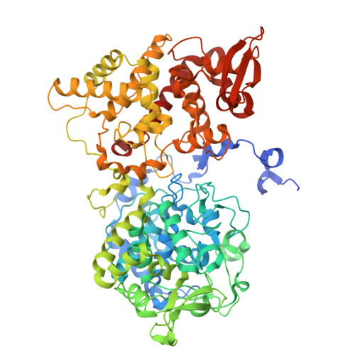
Catalase-peroxidase741Mycobacterium tuberculosisMutation(s): 0 
Gene Names: katG
EC: 1.11.1.21
UniProt
Find proteins for A0A0D5ZBI4 (Mycobacterium tuberculosis)
Explore A0A0D5ZBI4 
Go to UniProtKB:  A0A0D5ZBI4
Entity Groups  
Sequence Clusters30% Identity50% Identity70% Identity90% Identity95% Identity100% Identity
UniProt GroupA0A0D5ZBI4
Sequence Annotations
Expand
  • Reference Sequence
Find similar proteins by:  (by identity cutoff)  |  3D Structure
Entity ID: 2
MoleculeChains Sequence LengthOrganismDetailsImage
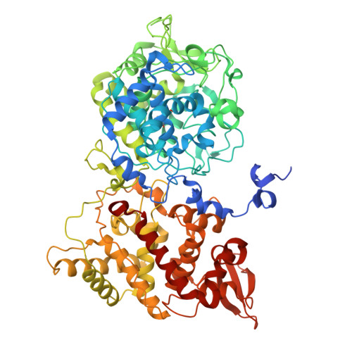
Catalase-peroxidase741Mycobacterium tuberculosisMutation(s): 0 
Gene Names: katGERS007661_00994ERS053720_01149
EC: 1.11.1.21
UniProt
Find proteins for A0A0D5ZBI4 (Mycobacterium tuberculosis)
Explore A0A0D5ZBI4 
Go to UniProtKB:  A0A0D5ZBI4
Entity Groups  
Sequence Clusters30% Identity50% Identity70% Identity90% Identity95% Identity100% Identity
UniProt GroupA0A0D5ZBI4
Sequence Annotations
Expand
  • Reference Sequence
Small Molecules
Ligands 5 Unique
IDChains Name / Formula / InChI Key2D Diagram3D Interactions
HEM (Subject of Investigation/LOI)
Query on HEM

Download Ideal Coordinates CCD File 
C [auth A],
G [auth B]
PROTOPORPHYRIN IX CONTAINING FE
C34 H32 Fe N4 O4
KABFMIBPWCXCRK-RGGAHWMASA-L
GOL
Query on GOL

Download Ideal Coordinates CCD File 
H [auth B],
I [auth B]
GLYCEROL
C3 H8 O3
PEDCQBHIVMGVHV-UHFFFAOYSA-N
ACT
Query on ACT

Download Ideal Coordinates CCD File 
F [auth A],
J [auth B]
ACETATE ION
C2 H3 O2
QTBSBXVTEAMEQO-UHFFFAOYSA-M
OXY (Subject of Investigation/LOI)
Query on OXY

Download Ideal Coordinates CCD File 
D [auth A]OXYGEN MOLECULE
O2
MYMOFIZGZYHOMD-UHFFFAOYSA-N
NA
Query on NA

Download Ideal Coordinates CCD File 
E [auth A]SODIUM ION
Na
FKNQFGJONOIPTF-UHFFFAOYSA-N
Modified Residues  1 Unique
IDChains TypeFormula2D DiagramParent
TOX
Query on TOX
B
L-PEPTIDE LINKINGC11 H12 N2 O4TRP
Experimental Data & Validation

Experimental Data

  • Method: X-RAY DIFFRACTION
  • Resolution: 2.35 Å
  • R-Value Free: 0.256 
  • R-Value Work: 0.191 
  • R-Value Observed: 0.193 
  • Space Group: P 42 21 2
Unit Cell:
Length ( Å )Angle ( ˚ )
a = 150.463α = 90
b = 150.463β = 90
c = 155.166γ = 90
Software Package:
Software NamePurpose
PHENIXrefinement
DENZOdata reduction
SCALEPACKdata scaling
PDB_EXTRACTdata extraction
PHASERphasing

Structure Validation

View Full Validation Report


Ligand Structure Quality Assessment 


Entry History & Funding Information

Deposition Data

  • Released Date: 2024-10-23 
  • Deposition Author(s): Liu, A., Li, J.

Funding OrganizationLocationGrant Number
National Institutes of Health/National Institute of General Medical Sciences (NIH/NIGMS)United StatesR01GM108988

Revision History  (Full details and data files)

  • Version 1.0: 2024-10-23
    Type: Initial release
  • Version 1.1: 2024-12-11
    Changes: Database references