


Crystal structure of S-adenosyl-L-homocysteine hydrolase from P. aeruginosa in complex with fragment F2X-Entry H09
Malecki, P.H., Gawel, M., Stepniewska, M., Brzezinski, K.To be published.
Experimental Data Snapshot
Starting Model: experimental
View more details
Entity ID: 1 | |||||
|---|---|---|---|---|---|
| Molecule | Chains | Sequence Length | Organism | Details | Image |
| Adenosylhomocysteinase | 472 | Pseudomonas aeruginosa PAO1 | Mutation(s): 0 Gene Names: ahcY, sahH, PA0432 EC: 3.3.1.1 (PDB Primary Data), 3.13.2.1 (UniProt) | 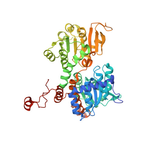 | |
UniProt | |||||
Find proteins for Q9I685 (Pseudomonas aeruginosa (strain ATCC 15692 / DSM 22644 / CIP 104116 / JCM 14847 / LMG 12228 / 1C / PRS 101 / PAO1)) Explore Q9I685 Go to UniProtKB: Q9I685 | |||||
Entity Groups | |||||
| Sequence Clusters | 30% Identity50% Identity70% Identity90% Identity95% Identity100% Identity | ||||
| UniProt Group | Q9I685 | ||||
Sequence AnnotationsExpand | |||||
| |||||
| Ligands 6 Unique | |||||
|---|---|---|---|---|---|
| ID | Chains | Name / Formula / InChI Key | 2D Diagram | 3D Interactions | |
| NAD Query on NAD | AB [auth J] EA [auth D] I [auth A] MA [auth H] MB [auth K] | NICOTINAMIDE-ADENINE-DINUCLEOTIDE C21 H27 N7 O14 P2 BAWFJGJZGIEFAR-NNYOXOHSSA-N |  | ||
| A1H8H (Subject of Investigation/LOI) Query on A1H8H | LA [auth D], LB [auth K], W [auth C], ZA [auth I] | 1-[2,4-bis(fluoranyl)phenyl]-2-(3,4-dihydro-1,2,4-triazol-2-yl)ethanone C10 H9 F2 N3 O UWNAFSDCDMGFTR-UHFFFAOYSA-N |  | ||
| ADE Query on ADE | BB [auth J] FA [auth D] J [auth A] NA [auth H] NB [auth K] | ADENINE C5 H5 N5 GFFGJBXGBJISGV-UHFFFAOYSA-N |  | ||
| PO4 Query on PO4 | AA [auth C] BA [auth C] CA [auth C] GA [auth D] GB [auth J] | PHOSPHATE ION O4 P NBIIXXVUZAFLBC-UHFFFAOYSA-K |  | ||
| GOL Query on GOL | CB [auth J], DB [auth J], EB [auth J], FB [auth J] | GLYCEROL C3 H8 O3 PEDCQBHIVMGVHV-UHFFFAOYSA-N |  | ||
| K Query on K | DA [auth C] KA [auth D] KB [auth J] O [auth A] RB [auth K] | POTASSIUM ION K NPYPAHLBTDXSSS-UHFFFAOYSA-N |  | ||
| Length ( Å ) | Angle ( ˚ ) |
|---|---|
| a = 111.073 | α = 90 |
| b = 210.799 | β = 105.835 |
| c = 111.501 | γ = 90 |
| Software Name | Purpose |
|---|---|
| PHENIX | refinement |
| XDS | data scaling |
| XDS | data reduction |
| REFMAC | phasing |
| Funding Organization | Location | Grant Number |
|---|---|---|
| Polish National Science Centre | Poland | SONATA BIS 2018/30/E/NZ1/00729 |
RCSB PDB is hosted by
RCSB PDB is a member of the

