8D4N

Crystal Structure of SARS-CoV-2 Main Protease (Mpro) E166Q Mutant


Experimental Data Snapshot

  • Method: X-RAY DIFFRACTION
  • Resolution: 2.70 Å
  • R-Value Free: 0.258 
  • R-Value Work: 0.206 
  • R-Value Observed: 0.209 

wwPDB Validation   3D Report Full Report


This is version 1.2 of the entry. See complete history


Literature

Naturally occurring mutations of SARS-CoV-2 main protease confer drug resistance to nirmatrelvir.

Hu, Y.Lewandowski, E.M.Tan, H.Zhang, X.Morgan, R.T.Zhang, X.Jacobs, L.M.C.Butler, S.G.Gongora, M.V.Choy, J.Deng, X.Chen, Y.Wang, J.

(2022) Biorxiv 

  • DOI: https://doi.org/10.1101/2022.06.28.497978
  • Primary Citation of Related Structures:  
    8D4J, 8D4K, 8D4L, 8D4M, 8D4N

  • PubMed Abstract: 

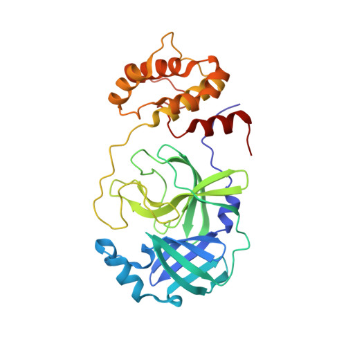
    The SARS-CoV-2 main protease (M pro ) is the drug target of Pfizer’s oral drug Paxlovid. The emergence of SARS-CoV-2 variants with mutations in M pro raised the alarm of potential drug resistance. In this study, we identified 100 naturally occurring M pro mutations located at the nirmatrelvir binding site, among which 20 mutants, including S144M/F/A/G/Y, M165T, E166G, H172Q/F, and Q192T/S/L/A/I/P/H/V/W/C/F, showed comparable enzymatic activity to the wild-type (k cat /K m <10-fold change) and resistance to nirmatrelvir (K i >10-fold increase). X-ray crystal structures were determined for seven representative mutants with and/or without GC-376/nirmatrelvir. Viral growth assay showed that M pro mutants with reduced enzymatic activity led to attenuated viral replication. Overall, our study identified several drug resistant hot spots that warrant close monitoring for possible clinical evidence of Paxlovid resistance.


  • Organizational Affiliation

    Department of Medicinal Chemistry, Ernest Mario School of Pharmacy, Rutgers, the State University of New Jersey, NJ, 08854, United States.


Macromolecules
Find similar proteins by:  (by identity cutoff)  |  3D Structure
Entity ID: 1
MoleculeChains Sequence LengthOrganismDetailsImage
3C-like proteinase nsp5
A, B
306Severe acute respiratory syndrome coronavirus 2Mutation(s): 1 
Gene Names: rep1a-1b
EC: 3.4.22.69
UniProt
Find proteins for P0DTD1 (Severe acute respiratory syndrome coronavirus 2)
Explore P0DTD1 
Go to UniProtKB:  P0DTD1
Entity Groups  
Sequence Clusters30% Identity50% Identity70% Identity90% Identity95% Identity100% Identity
UniProt GroupP0DTD1
Sequence Annotations
Expand
  • Reference Sequence
Experimental Data & Validation

Experimental Data

  • Method: X-RAY DIFFRACTION
  • Resolution: 2.70 Å
  • R-Value Free: 0.258 
  • R-Value Work: 0.206 
  • R-Value Observed: 0.209 
  • Space Group: P 1 21 1
Unit Cell:
Length ( Å )Angle ( ˚ )
a = 44.816α = 90
b = 53.687β = 101.79
c = 114.532γ = 90
Software Package:
Software NamePurpose
REFMACrefinement
PDB_EXTRACTdata extraction
HKL-2000data reduction
HKL-2000data scaling
PHASERphasing

Structure Validation

View Full Validation Report


Entry History & Funding Information

Deposition Data


Funding OrganizationLocationGrant Number
National Institutes of Health/National Institute Of Allergy and Infectious Diseases (NIH/NIAID)United StatesAI158775

Revision History  (Full details and data files)

  • Version 1.0: 2022-07-13
    Type: Initial release
  • Version 1.1: 2022-10-05
    Changes: Database references
  • Version 1.2: 2023-10-18
    Changes: Data collection, Refinement description