8DVH

Crystal structure of ATP-dependent Lon protease from Bacillus subtillis (BsLonBA)


Experimental Data Snapshot

  • Method: X-RAY DIFFRACTION
  • Resolution: 1.90 Å
  • R-Value Free: 0.237 
  • R-Value Work: 0.194 
  • R-Value Observed: 0.195 

wwPDB Validation   3D Report Full Report


Ligand Structure Quality Assessment 


This is version 1.1 of the entry. See complete history


Literature

Unique Structural Fold of LonBA Protease from Bacillus subtilis, a Member of a Newly Identified Subfamily of Lon Proteases.

Gustchina, A.Li, M.Andrianova, A.G.Kudzhaev, A.M.Lountos, G.T.Sekula, B.Cherry, S.Tropea, J.E.Smirnov, I.V.Wlodawer, A.Rotanova, T.V.

(2022) Int J Mol Sci 23

  • DOI: https://doi.org/10.3390/ijms231911425
  • Primary Citation of Related Structures:  
    8DVH

  • PubMed Abstract: 

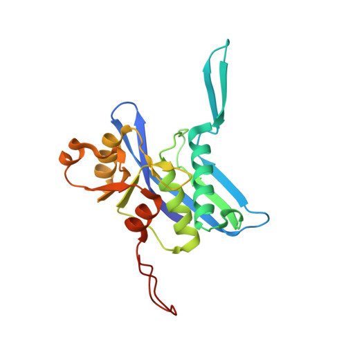
    ATP-dependent Lon proteases are key participants in the quality control system that supports the homeostasis of the cellular proteome. Based on their unique structural and biochemical properties, Lon proteases have been assigned in the MEROPS database to three subfamilies (A, B, and C). All Lons are single-chain, multidomain proteins containing an ATPase and protease domains, with different additional elements present in each subfamily. LonA and LonC proteases are soluble cytoplasmic enzymes, whereas LonBs are membrane-bound. Based on an analysis of the available sequences of Lon proteases, we identified a number of enzymes currently assigned to the LonB subfamily that, although presumably membrane-bound, include structural features more similar to their counterparts in the LonA subfamily. This observation was confirmed by the crystal structure of the proteolytic domain of the enzyme previously assigned as Bacillus subtilis LonB, combined with the modeled structure of its ATPase domain. Several structural features present in both domains differ from their counterparts in either LonA or LonB subfamilies. We thus postulate that this enzyme is the founding member of a newly identified LonBA subfamily, so far found only in the gene sequences of firmicutes.


  • Organizational Affiliation

    Center for Structural Biology, National Cancer Institute, Frederick, MD 21702, USA.


Macromolecules
Find similar proteins by:  (by identity cutoff)  |  3D Structure
Entity ID: 1
MoleculeChains Sequence LengthOrganismDetailsImage
Lon protease 2
A, B
206Bacillus subtilis subsp. subtilis str. 168Mutation(s): 0 
Gene Names: lon2lonBysxFBSU28210
EC: 3.4.21.53
UniProt
Find proteins for P42425 (Bacillus subtilis (strain 168))
Explore P42425 
Go to UniProtKB:  P42425
Entity Groups  
Sequence Clusters30% Identity50% Identity70% Identity90% Identity95% Identity100% Identity
UniProt GroupP42425
Sequence Annotations
Expand
  • Reference Sequence
Experimental Data & Validation

Experimental Data

  • Method: X-RAY DIFFRACTION
  • Resolution: 1.90 Å
  • R-Value Free: 0.237 
  • R-Value Work: 0.194 
  • R-Value Observed: 0.195 
  • Space Group: P 63
Unit Cell:
Length ( Å )Angle ( ˚ )
a = 89.892α = 90
b = 89.892β = 90
c = 83.95γ = 120
Software Package:
Software NamePurpose
PDB_EXTRACTdata extraction
REFMACrefinement
PHENIXmodel building
PHASERphasing
Aimlessdata scaling
XDSdata reduction

Structure Validation

View Full Validation Report


Ligand Structure Quality Assessment 


Entry History & Funding Information

Deposition Data


Funding OrganizationLocationGrant Number
National Institutes of Health/National Cancer Institute (NIH/NCI)United StatesIntramural Research Program
Russian Science FoundationRussian FederationRussian Science Foundation

Revision History  (Full details and data files)

  • Version 1.0: 2022-11-09
    Type: Initial release
  • Version 1.1: 2023-10-18
    Changes: Data collection, Refinement description