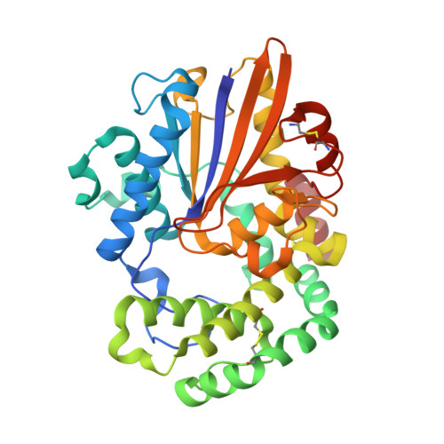
8GDL

Acid phosphatase pseudoenzyme from flea


Experimental Data Snapshot

  • Method: X-RAY DIFFRACTION
  • Resolution: 1.75 Å
  • R-Value Free: 0.250 
  • R-Value Work: 0.215 
  • R-Value Observed: 0.217 

Starting Model: in silico
View more details

wwPDB Validation   3D Report Full Report


Ligand Structure Quality Assessment 


This is version 1.1 of the entry. See complete history


Literature

Acid phosphatase-like proteins, a biogenic amine and leukotriene-binding salivary protein family from the flea Xenopsylla cheopis.

Lu, S.Andersen, J.F.Bosio, C.F.Hinnebusch, B.J.Ribeiro, J.M.

(2023) Commun Biol 6: 1280-1280

  • DOI: https://doi.org/10.1038/s42003-023-05679-0
  • Primary Citation of Related Structures:  
    8GDL

  • PubMed Abstract: 

    The salivary glands of hematophagous arthropods contain pharmacologically active molecules that interfere with host hemostasis and immune responses, favoring blood acquisition and pathogen transmission. Exploration of the salivary gland composition of the rat flea, Xenopsylla cheopis, revealed several abundant acid phosphatase-like proteins whose sequences lacked one or two of their presumed catalytic residues. In this study, we undertook a comprehensive characterization of the tree most abundant X. cheopis salivary acid phosphatase-like proteins. Our findings indicate that the three recombinant proteins lacked the anticipated catalytic activity and instead, displayed the ability to bind different biogenic amines and leukotrienes with high affinity. Moreover, X-ray crystallography data from the XcAP-1 complexed with serotonin revealed insights into their binding mechanisms.


  • Organizational Affiliation

    Laboratory of Malaria and Vector Research, National Institute of Allergy and Infectious Diseases, Bethesda, MD, USA. Stephen.lu@nih.gov.


Macromolecules
Find similar proteins by:  (by identity cutoff)  |  3D Structure
Entity ID: 1
MoleculeChains Sequence LengthOrganismDetailsImage
Secreted salivary acid phosphatase
A, B
361Xenopsylla cheopisMutation(s): 0 
UniProt
Find proteins for A2IA89 (Xenopsylla cheopis)
Explore A2IA89 
Go to UniProtKB:  A2IA89
Entity Groups  
Sequence Clusters30% Identity50% Identity70% Identity90% Identity95% Identity100% Identity
UniProt GroupA2IA89
Sequence Annotations
Expand
  • Reference Sequence
Small Molecules
Ligands 3 Unique
IDChains Name / Formula / InChI Key2D Diagram3D Interactions
PAM
Query on PAM

Download Ideal Coordinates CCD File 
G [auth A],
J [auth B]
PALMITOLEIC ACID
C16 H30 O2
SECPZKHBENQXJG-FPLPWBNLSA-N
SRO
Query on SRO

Download Ideal Coordinates CCD File 
C [auth A],
H [auth B]
SEROTONIN
C10 H12 N2 O
QZAYGJVTTNCVMB-UHFFFAOYSA-N
SO4
Query on SO4

Download Ideal Coordinates CCD File 
D [auth A],
E [auth A],
F [auth A],
I [auth B]
SULFATE ION
O4 S
QAOWNCQODCNURD-UHFFFAOYSA-L
Experimental Data & Validation

Experimental Data

  • Method: X-RAY DIFFRACTION
  • Resolution: 1.75 Å
  • R-Value Free: 0.250 
  • R-Value Work: 0.215 
  • R-Value Observed: 0.217 
  • Space Group: P 1 21 1
Unit Cell:
Length ( Å )Angle ( ˚ )
a = 47.968α = 90
b = 71.898β = 91.54
c = 113.513γ = 90
Software Package:
Software NamePurpose
PHENIXrefinement
HKL-2000data scaling
HKL-2000data reduction
PHASERphasing

Structure Validation

View Full Validation Report


Ligand Structure Quality Assessment 


Entry History & Funding Information

Deposition Data


Funding OrganizationLocationGrant Number
National Institutes of Health/National Institute Of Allergy and Infectious Diseases (NIH/NIAID)United States--

Revision History  (Full details and data files)

  • Version 1.0: 2024-01-17
    Type: Initial release
  • Version 1.1: 2024-10-23
    Changes: Structure summary