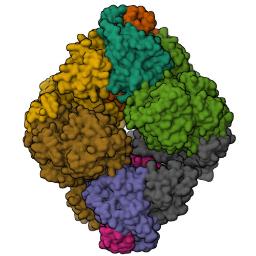
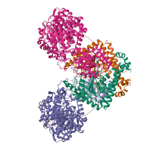
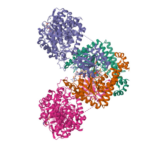
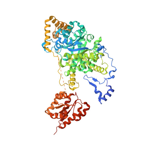
8GJU

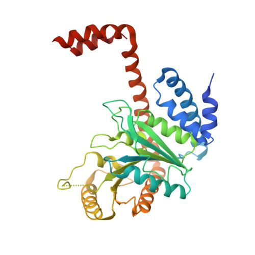
Crystal structure of human methylmalonyl-CoA mutase (MMUT) in complex with methylmalonic acidemia type A protein (MMAA), coenzyme A, and GDP


Experimental Data Snapshot

  • Method: X-RAY DIFFRACTION
  • Resolution: 2.79 Å
  • R-Value Free: 0.276 
  • R-Value Work: 0.229 
  • R-Value Observed: 0.232 

Starting Models: experimental
View more details

wwPDB Validation   3D Report Full Report


Ligand Structure Quality Assessment 


This is version 1.1 of the entry. See complete history


Literature

Architecture of the human G-protein-methylmalonyl-CoA mutase nanoassembly for B 12 delivery and repair.

Mascarenhas, R.Ruetz, M.Gouda, H.Heitman, N.Yaw, M.Banerjee, R.

(2023) Nat Commun 14: 4332-4332

  • DOI: https://doi.org/10.1038/s41467-023-40077-4
  • Primary Citation of Related Structures:  
    8GJU

  • PubMed Abstract: 

    G-proteins function as molecular switches to power cofactor translocation and confer fidelity in metal trafficking. The G-protein, MMAA, together with MMAB, an adenosyltransferase, orchestrate cofactor delivery and repair of B 12 -dependent human methylmalonyl-CoA mutase (MMUT). The mechanism by which the complex assembles and moves a >1300 Da cargo, or fails in disease, are poorly understood. Herein, we report the crystal structure of the human MMUT-MMAA nano-assembly, which reveals a dramatic 180° rotation of the B 12 domain, exposing it to solvent. The complex, stabilized by MMAA wedging between two MMUT domains, leads to ordering of the switch I and III loops, revealing the molecular basis of mutase-dependent GTPase activation. The structure explains the biochemical penalties incurred by methylmalonic aciduria-causing mutations that reside at the MMAA-MMUT interfaces we identify here.


  • Organizational Affiliation

    Department of Biological Chemistry, University of Michigan, Ann Arbor, MI, 48109, USA.


Macromolecules
Find similar proteins by:  (by identity cutoff)  |  3D Structure
Entity ID: 1
MoleculeChains Sequence LengthOrganismDetailsImage
Methylmalonic aciduria type A protein, mitochondrialA [auth D],
B [auth F],
F [auth A],
G [auth B]
349Homo sapiensMutation(s): 0 
Gene Names: MMAA
EC: 3.6
UniProt & NIH Common Fund Data Resources
Find proteins for Q8IVH4 (Homo sapiens)
Explore Q8IVH4 
Go to UniProtKB:  Q8IVH4
PHAROS:  Q8IVH4
GTEx:  ENSG00000151611 
Entity Groups  
Sequence Clusters30% Identity50% Identity70% Identity90% Identity95% Identity100% Identity
UniProt GroupQ8IVH4
Sequence Annotations
Expand
  • Reference Sequence
Find similar proteins by:  (by identity cutoff)  |  3D Structure
Entity ID: 2
MoleculeChains Sequence LengthOrganismDetailsImage
Methylmalonyl-CoA mutase, mitochondrialC [auth J],
D [auth K],
E [auth L],
H
748Homo sapiensMutation(s): 0 
Gene Names: MUT
EC: 5.4.99.2
UniProt & NIH Common Fund Data Resources
Find proteins for P22033 (Homo sapiens)
Explore P22033 
Go to UniProtKB:  P22033
PHAROS:  P22033
GTEx:  ENSG00000146085 
Entity Groups  
Sequence Clusters30% Identity50% Identity70% Identity90% Identity95% Identity100% Identity
UniProt GroupP22033
Sequence Annotations
Expand
  • Reference Sequence
Small Molecules
Ligands 3 Unique
IDChains Name / Formula / InChI Key2D Diagram3D Interactions
COA (Subject of Investigation/LOI)
Query on COA

Download Ideal Coordinates CCD File 
M [auth J],
N [auth K],
O [auth L],
T [auth H]
COENZYME A
C21 H36 N7 O16 P3 S
RGJOEKWQDUBAIZ-IBOSZNHHSA-N
GDP (Subject of Investigation/LOI)
Query on GDP

Download Ideal Coordinates CCD File 
I [auth D],
K [auth F],
P [auth A],
R [auth B]
GUANOSINE-5'-DIPHOSPHATE
C10 H15 N5 O11 P2
QGWNDRXFNXRZMB-UUOKFMHZSA-N
MG
Query on MG

Download Ideal Coordinates CCD File 
J [auth D],
L [auth F],
Q [auth A],
S [auth B]
MAGNESIUM ION
Mg
JLVVSXFLKOJNIY-UHFFFAOYSA-N
Experimental Data & Validation

Experimental Data

  • Method: X-RAY DIFFRACTION
  • Resolution: 2.79 Å
  • R-Value Free: 0.276 
  • R-Value Work: 0.229 
  • R-Value Observed: 0.232 
  • Space Group: P 1 21 1
Unit Cell:
Length ( Å )Angle ( ˚ )
a = 95.806α = 90
b = 221.872β = 105.53
c = 121.824γ = 90
Software Package:
Software NamePurpose
PHENIXrefinement
XDSdata reduction
XDSdata scaling
PHASERphasing

Structure Validation

View Full Validation Report


Ligand Structure Quality Assessment 


Entry History & Funding Information

Deposition Data


Funding OrganizationLocationGrant Number
National Institutes of Health/National Institute of Diabetes and Digestive and Kidney Disease (NIH/NIDDK)United StatesDK45776
National Institutes of Health/National Institute of General Medical Sciences (NIH/NIGMS)United StatesK99-GM1434820

Revision History  (Full details and data files)

  • Version 1.0: 2023-08-09
    Type: Initial release
  • Version 1.1: 2024-05-22
    Changes: Data collection