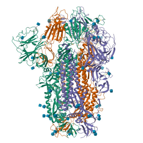
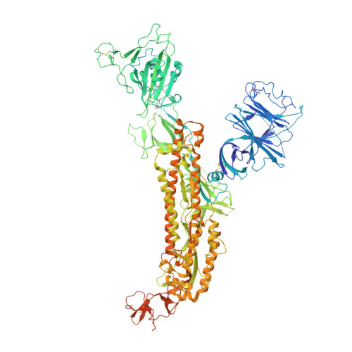
8IOT

Structure of the SARS-CoV-2 XBB.1 spike glycoprotein (closed-2 state)


Experimental Data Snapshot

  • Method: ELECTRON MICROSCOPY
  • Resolution: 2.51 Å
  • Aggregation State: PARTICLE 
  • Reconstruction Method: SINGLE PARTICLE 

Starting Model: experimental
View more details

wwPDB Validation   3D Report Full Report


This is version 1.2 of the entry. See complete history


Literature

Virological characteristics of the SARS-CoV-2 XBB variant derived from recombination of two Omicron subvariants.

Tamura, T.Ito, J.Uriu, K.Zahradnik, J.Kida, I.Anraku, Y.Nasser, H.Shofa, M.Oda, Y.Lytras, S.Nao, N.Itakura, Y.Deguchi, S.Suzuki, R.Wang, L.Begum, M.M.Kita, S.Yajima, H.Sasaki, J.Sasaki-Tabata, K.Shimizu, R.Tsuda, M.Kosugi, Y.Fujita, S.Pan, L.Sauter, D.Yoshimatsu, K.Suzuki, S.Asakura, H.Nagashima, M.Sadamasu, K.Yoshimura, K.Yamamoto, Y.Nagamoto, T.Schreiber, G.Maenaka, K.Hashiguchi, T.Ikeda, T.Fukuhara, T.Saito, A.Tanaka, S.Matsuno, K.Takayama, K.Sato, K.

(2023) Nat Commun 14: 2800-2800

  • DOI: https://doi.org/10.1038/s41467-023-38435-3
  • Primary Citation of Related Structures:  
    8IOS, 8IOT, 8IOU, 8IOV

  • PubMed Abstract: 

    In late 2022, SARS-CoV-2 Omicron subvariants have become highly diversified, and XBB is spreading rapidly around the world. Our phylogenetic analyses suggested that XBB emerged through the recombination of two cocirculating BA.2 lineages, BJ.1 and BM.1.1.1 (a progeny of BA.2.75), during the summer of 2022. XBB.1 is the variant most profoundly resistant to BA.2/5 breakthrough infection sera to date and is more fusogenic than BA.2.75. The recombination breakpoint is located in the receptor-binding domain of spike, and each region of the recombinant spike confers immune evasion and increases fusogenicity. We further provide the structural basis for the interaction between XBB.1 spike and human ACE2. Finally, the intrinsic pathogenicity of XBB.1 in male hamsters is comparable to or even lower than that of BA.2.75. Our multiscale investigation provides evidence suggesting that XBB is the first observed SARS-CoV-2 variant to increase its fitness through recombination rather than substitutions.


  • Organizational Affiliation

    Department of Microbiology and Immunology, Faculty of Medicine, Hokkaido University, Sapporo, Japan.


Macromolecules
Find similar proteins by:  (by identity cutoff)  |  3D Structure
Entity ID: 1
MoleculeChains Sequence LengthOrganismDetailsImage
Spike glycoprotein
A, B, C
1,245Severe acute respiratory syndrome coronavirus 2Mutation(s): 47 
Gene Names: S2
UniProt
Find proteins for P0DTC2 (Severe acute respiratory syndrome coronavirus 2)
Explore P0DTC2 
Go to UniProtKB:  P0DTC2
Entity Groups  
Sequence Clusters30% Identity50% Identity70% Identity90% Identity95% Identity100% Identity
UniProt GroupP0DTC2
Glycosylation
Glycosylation Sites: 14Go to GlyGen: P0DTC2-1
Sequence Annotations
Expand
  • Reference Sequence
Oligosaccharides

Help

Entity ID: 2
MoleculeChains Length2D Diagram Glycosylation3D Interactions
2-acetamido-2-deoxy-beta-D-glucopyranose-(1-4)-2-acetamido-2-deoxy-beta-D-glucopyranose
D, E, F, G, H
D, E, F, G, H, I
2N-Glycosylation
Glycosylation Resources
GlyTouCan:  G42666HT
GlyCosmos:  G42666HT
GlyGen:  G42666HT
Small Molecules
Ligands 1 Unique
IDChains Name / Formula / InChI Key2D Diagram3D Interactions
NAG
Query on NAG

Download Ideal Coordinates CCD File 
AA [auth B]
BA [auth B]
CA [auth B]
DA [auth B]
EA [auth B]
AA [auth B],
BA [auth B],
CA [auth B],
DA [auth B],
EA [auth B],
FA [auth B],
GA [auth B],
HA [auth C],
IA [auth C],
J [auth A],
JA [auth C],
K [auth A],
KA [auth C],
L [auth A],
LA [auth C],
M [auth A],
MA [auth C],
N [auth A],
NA [auth C],
O [auth A],
OA [auth C],
P [auth A],
PA [auth C],
Q [auth A],
QA [auth C],
R [auth A],
RA [auth C],
S [auth A],
SA [auth C],
T [auth A],
U [auth A],
V [auth B],
W [auth B],
X [auth B],
Y [auth B],
Z [auth B]
2-acetamido-2-deoxy-beta-D-glucopyranose
C8 H15 N O6
OVRNDRQMDRJTHS-FMDGEEDCSA-N
Experimental Data & Validation

Experimental Data

  • Method: ELECTRON MICROSCOPY
  • Resolution: 2.51 Å
  • Aggregation State: PARTICLE 
  • Reconstruction Method: SINGLE PARTICLE 
EM Software:
TaskSoftware PackageVersion
MODEL REFINEMENTPHENIX1.20
RECONSTRUCTIONcryoSPARC4.1.2

Structure Validation

View Full Validation Report


Entry History & Funding Information

Deposition Data


Funding OrganizationLocationGrant Number
Japan Agency for Medical Research and Development (AMED)JapanJP21am0101093
Japan Agency for Medical Research and Development (AMED)JapanJP22ama121037
Japan Science and TechnologyJapanJPMJCR20H8
Japan Society for the Promotion of Science (JSPS)JapanJPJSCCA20190008
Japan Society for the Promotion of Science (JSPS)Japan20H05773
Japan Society for the Promotion of Science (JSPS)JapanJP20H05873

Revision History  (Full details and data files)

  • Version 1.0: 2023-05-24
    Type: Initial release
  • Version 1.1: 2023-07-19
    Changes: Database references
  • Version 1.2: 2024-10-30
    Changes: Data collection, Refinement description, Structure summary