


Aspartyl/Asparaginyl beta-hydroxylase (AspH) in complex with Mn, 2-oxoglutarate and a Factor X derived peptide fragment
Brasnett, A., Hou, C., Rabe, P., Brewitz, L., Schofield, C.J.To be published.
Experimental Data Snapshot
Starting Model: experimental
View more details
Entity ID: 1 | |||||
|---|---|---|---|---|---|
| Molecule | Chains | Sequence Length | Organism | Details | Image |
| Aspartyl/asparaginyl beta-hydroxylase | 444 | Homo sapiens | Mutation(s): 0 Gene Names: ASPH, BAH EC: 1.14.11.16 | 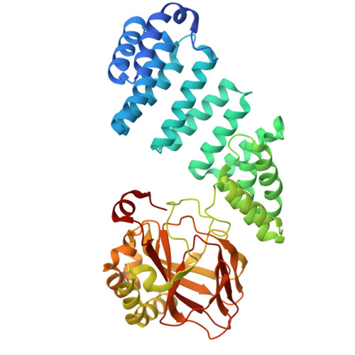 | |
UniProt & NIH Common Fund Data Resources | |||||
Find proteins for Q12797 (Homo sapiens) Explore Q12797 Go to UniProtKB: Q12797 | |||||
PHAROS: Q12797 GTEx: ENSG00000198363 | |||||
Entity Groups | |||||
| Sequence Clusters | 30% Identity50% Identity70% Identity90% Identity95% Identity100% Identity | ||||
| UniProt Group | Q12797 | ||||
Sequence AnnotationsExpand | |||||
| |||||
Entity ID: 2 | |||||
|---|---|---|---|---|---|
| Molecule | Chains | Sequence Length | Organism | Details | Image |
| Coagulation factor X | 39 | Homo sapiens | Mutation(s): 4 Gene Names: F10 EC: 3.4.21.6 | 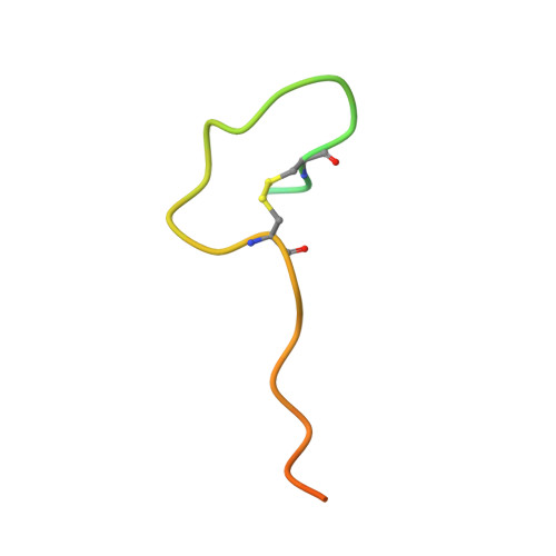 | |
UniProt & NIH Common Fund Data Resources | |||||
Find proteins for P00742 (Homo sapiens) Explore P00742 Go to UniProtKB: P00742 | |||||
PHAROS: P00742 GTEx: ENSG00000126218 | |||||
Entity Groups | |||||
| Sequence Clusters | 30% Identity50% Identity70% Identity90% Identity95% Identity100% Identity | ||||
| UniProt Group | P00742 | ||||
Sequence AnnotationsExpand | |||||
| |||||
| Ligands 3 Unique | |||||
|---|---|---|---|---|---|
| ID | Chains | Name / Formula / InChI Key | 2D Diagram | 3D Interactions | |
| AKG (Subject of Investigation/LOI) Query on AKG | C [auth A] | 2-OXOGLUTARIC ACID C5 H6 O5 KPGXRSRHYNQIFN-UHFFFAOYSA-N |  | ||
| PEG Query on PEG | E [auth A] F [auth A] G [auth A] H [auth A] I [auth A] | DI(HYDROXYETHYL)ETHER C4 H10 O3 MTHSVFCYNBDYFN-UHFFFAOYSA-N |  | ||
| MN (Subject of Investigation/LOI) Query on MN | D [auth A] | MANGANESE (II) ION Mn WAEMQWOKJMHJLA-UHFFFAOYSA-N |  | ||
| Length ( Å ) | Angle ( ˚ ) |
|---|---|
| a = 49.68 | α = 90 |
| b = 86.19 | β = 90 |
| c = 123.35 | γ = 90 |
| Software Name | Purpose |
|---|---|
| PHENIX | refinement |
| xia2 | data reduction |
| xia2 | data scaling |
| PHASER | phasing |
| Funding Organization | Location | Grant Number |
|---|---|---|
| Wellcome Trust | United Kingdom | 106244/Z/14/Z |
| Biotechnology and Biological Sciences Research Council (BBSRC) | United Kingdom | BB/V001892/1 |
| Wellcome Trust | United Kingdom | 227298/Z/23/Z |
RCSB PDB is hosted by

RCSB PDB is a member of the

All questions and comments will receive a response in a timely manner.

