8ST4

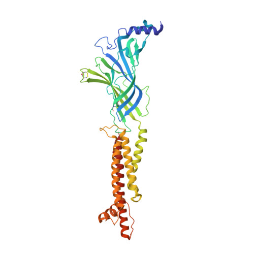
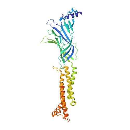
The 2alpha3beta stoichiometry of human alpha4beta2 nicotinic acetylcholine receptor in complex with acetylcholine

  • Classification: TRANSPORT PROTEIN
  • Organism(s): Homo sapiens, Mus musculus
  • Expression System: Homo sapiens
  • Mutation(s): No 
  • Membrane Protein: Yes  PDBTMMemProtMD

  • Deposited: 2023-05-09 Released: 2024-02-07 
  • Deposition Author(s): Kang, G., Hibbs, R.E.
  • Funding Organization(s): National Institutes of Health/National Institute on Drug Abuse (NIH/NIDA), National Institutes of Health/National Institute of Neurological Disorders and Stroke (NIH/NINDS)

Experimental Data Snapshot

  • Method: ELECTRON MICROSCOPY
  • Resolution: 2.35 Å
  • Aggregation State: PARTICLE 
  • Reconstruction Method: SINGLE PARTICLE 

wwPDB Validation   3D Report Full Report


This is version 1.2 of the entry. See complete history


Literature

Structural bases for stoichiometry-selective calcium potentiation of a neuronal nicotinic receptor.

Mazzaferro, S.Kang, G.Natarajan, K.Hibbs, R.E.Sine, S.M.

(2024) Br J Pharmacol 181: 1973-1992

  • DOI: https://doi.org/10.1111/bph.16321
  • Primary Citation of Related Structures:  
    8SSZ, 8ST0, 8ST1, 8ST2, 8ST3, 8ST4

  • PubMed Abstract: 

    α4β2 nicotinic acetylcholine (nACh) receptors assemble in two stoichiometric forms, one of which is potentiated by calcium. The sites of calcium binding that underpin potentiation are not known. To identify calcium binding sites, we applied cryo-electron microscopy (cryo-EM) and molecular dynamics (MD) simulations to each stoichiometric form of the α4β2 nACh receptor in the presence of calcium ions. To test whether the identified calcium sites are linked to potentiation, we generated mutants of anionic residues at the sites, expressed wild type and mutant receptors in clonal mammalian fibroblasts, and recorded ACh-elicited single-channel currents with or without calcium. Both cryo-EM and MD simulations show calcium bound to a site between the extracellular and transmembrane domains of each α4 subunit (ECD-TMD site). Substituting alanine for anionic residues at the ECD-TMD site abolishes stoichiometry-selective calcium potentiation, as monitored by single-channel patch clamp electrophysiology. Additionally, MD simulation reveals calcium association at subunit interfaces within the extracellular domain. Substituting alanine for anionic residues at the ECD sites reduces or abolishes stoichiometry-selective calcium potentiation. Stoichiometry-selective calcium potentiation of the α4β2 nACh receptor is achieved by calcium association with topographically distinct sites framed by anionic residues within the α4 subunit and between the α4 and β2 subunits. Stoichiometry-selective calcium potentiation could result from the greater number of calcium sites in the stoichiometric form with three rather than two α4 subunits. The results are relevant to modulation of signalling via α4β2 nACh receptors in physiological and pathophysiological conditions.


  • Organizational Affiliation

    Department of Physiology and Biomedical Engineering, Mayo Clinic, Rochester, Minnesota, USA.


Macromolecules
Find similar proteins by:  (by identity cutoff)  |  3D Structure
Entity ID: 1
MoleculeChains Sequence LengthOrganismDetailsImage
Neuronal acetylcholine receptor subunit alpha-4
A, D
386Homo sapiensMutation(s): 0 
Gene Names: CHRNA4NACRA4
Membrane Entity: Yes 
UniProt & NIH Common Fund Data Resources
Find proteins for P43681 (Homo sapiens)
Explore P43681 
Go to UniProtKB:  P43681
PHAROS:  P43681
GTEx:  ENSG00000101204 
Entity Groups  
Sequence Clusters30% Identity50% Identity70% Identity90% Identity95% Identity100% Identity
UniProt GroupP43681
Glycosylation
Glycosylation Sites: 3Go to GlyGen: P43681-1
Sequence Annotations
Expand
  • Reference Sequence
Find similar proteins by:  (by identity cutoff)  |  3D Structure
Entity ID: 2
MoleculeChains Sequence LengthOrganismDetailsImage
Neuronal acetylcholine receptor subunit beta-2
B, C, E
403Homo sapiensMutation(s): 0 
Gene Names: CHRNB2
Membrane Entity: Yes 
UniProt & NIH Common Fund Data Resources
Find proteins for P17787 (Homo sapiens)
Explore P17787 
Go to UniProtKB:  P17787
PHAROS:  P17787
GTEx:  ENSG00000160716 
Entity Groups  
Sequence Clusters30% Identity50% Identity70% Identity90% Identity95% Identity100% Identity
UniProt GroupP17787
Glycosylation
Glycosylation Sites: 1Go to GlyGen: P17787-1
Sequence Annotations
Expand
  • Reference Sequence
Find similar proteins by:  (by identity cutoff)  |  3D Structure
Entity ID: 3
MoleculeChains Sequence LengthOrganismDetailsImage
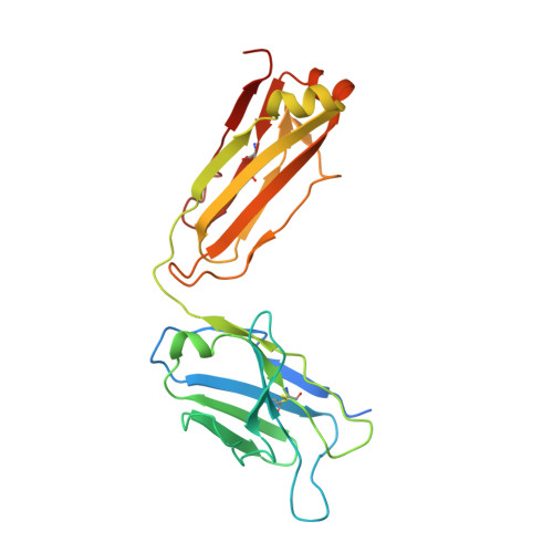
IgG1 Kappa Light Chain
F, H, J
238Mus musculusMutation(s): 0 
Entity Groups  
Sequence Clusters30% Identity50% Identity70% Identity90% Identity95% Identity100% Identity
Sequence Annotations
Expand
  • Reference Sequence
Find similar proteins by:  (by identity cutoff)  |  3D Structure
Entity ID: 4
MoleculeChains Sequence LengthOrganismDetailsImage
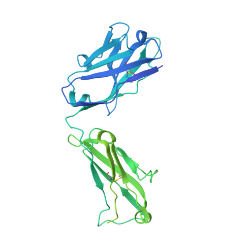
IgG1 Heavy Chain
G, I, K
462Mus musculusMutation(s): 0 
Entity Groups  
Sequence Clusters30% Identity50% Identity70% Identity90% Identity95% Identity100% Identity
Sequence Annotations
Expand
  • Reference Sequence
Oligosaccharides

Help

Entity ID: 5
MoleculeChains Length2D Diagram Glycosylation3D Interactions
beta-D-mannopyranose-(1-4)-2-acetamido-2-deoxy-beta-D-glucopyranose-(1-4)-2-acetamido-2-deoxy-beta-D-glucopyranose
L, M, N
3N-Glycosylation
Glycosylation Resources
GlyTouCan:  G15407YE
GlyCosmos:  G15407YE
GlyGen:  G15407YE
Small Molecules
Ligands 3 Unique
IDChains Name / Formula / InChI Key2D Diagram3D Interactions
NAG
Query on NAG

Download Ideal Coordinates CCD File 
O [auth A]
P [auth A]
Q [auth A]
T [auth D]
U [auth D]
O [auth A],
P [auth A],
Q [auth A],
T [auth D],
U [auth D],
V [auth D]
2-acetamido-2-deoxy-beta-D-glucopyranose
C8 H15 N O6
OVRNDRQMDRJTHS-FMDGEEDCSA-N
ACH (Subject of Investigation/LOI)
Query on ACH

Download Ideal Coordinates CCD File 
R [auth A],
W [auth D]
ACETYLCHOLINE
C7 H16 N O2
OIPILFWXSMYKGL-UHFFFAOYSA-N
NA
Query on NA

Download Ideal Coordinates CCD File 
S [auth B]SODIUM ION
Na
FKNQFGJONOIPTF-UHFFFAOYSA-N
Experimental Data & Validation

Experimental Data

  • Method: ELECTRON MICROSCOPY
  • Resolution: 2.35 Å
  • Aggregation State: PARTICLE 
  • Reconstruction Method: SINGLE PARTICLE 
EM Software:
TaskSoftware PackageVersion
MODEL REFINEMENTPHENIX1.20.1_4487

Structure Validation

View Full Validation Report


Entry History & Funding Information

Deposition Data


Funding OrganizationLocationGrant Number
National Institutes of Health/National Institute on Drug Abuse (NIH/NIDA)United StatesDA047848
National Institutes of Health/National Institute on Drug Abuse (NIH/NIDA)United StatesDA042072
National Institutes of Health/National Institute of Neurological Disorders and Stroke (NIH/NINDS)United StatesNS120496

Revision History  (Full details and data files)

  • Version 1.0: 2024-02-07
    Type: Initial release
  • Version 1.1: 2024-08-21
    Changes: Data collection, Database references
  • Version 1.2: 2024-11-06
    Changes: Data collection, Structure summary