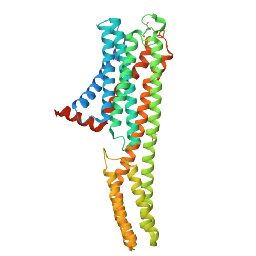
8TF5

Crystal structure of orphan G protein-coupled receptor 6, pseudoapo form


Experimental Data Snapshot

  • Method: X-RAY DIFFRACTION
  • Resolution: 2.10 Å
  • R-Value Free: 0.214 
  • R-Value Work: 0.184 
  • R-Value Observed: 0.186 

Starting Model: experimental
View more details

wwPDB Validation   3D Report Full Report


Ligand Structure Quality Assessment 


This is version 1.1 of the entry. See complete history


Literature

Structural insights into the high basal activity and inverse agonism of the orphan receptor GPR6 implicated in Parkinson's disease.

Barekatain, M.Johansson, L.C.Lam, J.H.Chang, H.Sadybekov, A.V.Han, G.W.Russo, J.Bliesath, J.Brice, N.L.Carlton, M.B.L.Saikatendu, K.S.Sun, H.Murphy, S.T.Monenschein, H.Schiffer, H.H.Popov, P.Lutomski, C.A.Robinson, C.V.Liu, Z.J.Hua, T.Katritch, V.Cherezov, V.

(2024) Sci Signal 17: eado8741-eado8741

  • DOI: https://doi.org/10.1126/scisignal.ado8741
  • Primary Citation of Related Structures:  
    8T1V, 8T1W, 8TF5, 8TYW

  • PubMed Abstract: 

    GPR6 is an orphan G protein-coupled receptor with high constitutive activity found in D2-type dopamine receptor-expressing medium spiny neurons of the striatopallidal pathway, which is aberrantly hyperactivated in Parkinson's disease. Here, we solved crystal structures of GPR6 without the addition of a ligand (a pseudo-apo state) and in complex with two inverse agonists, including CVN424, which improved motor symptoms in patients with Parkinson's disease in clinical trials. In addition, we obtained a cryo-electron microscopy structure of the signaling complex between GPR6 and its cognate G s heterotrimer. The pseudo-apo structure revealed a strong density in the orthosteric pocket of GPR6 corresponding to a lipid-like endogenous ligand. A combination of site-directed mutagenesis, native mass spectrometry, and computer modeling suggested potential mechanisms for high constitutive activity and inverse agonism in GPR6 and identified a series of lipids and ions bound to the receptor. The structures and results obtained in this study could guide the rational design of drugs that modulate GPR6 signaling.


  • Organizational Affiliation

    Bridge Institute, USC Michelson Center for Convergent Biosciences, University of Southern California, Los Angeles, CA 90089, USA.


Macromolecules
Find similar proteins by:  (by identity cutoff)  |  3D Structure
Entity ID: 1
MoleculeChains Sequence LengthOrganismDetailsImage
G-protein coupled receptor 6,Soluble cytochrome b562 chimera,Soluble cytochrome b562,G-protein coupled receptor 6436Homo sapiensEscherichia coliMutation(s): 9 
Gene Names: GPR6cybC
Membrane Entity: Yes 
UniProt & NIH Common Fund Data Resources
Find proteins for P46095 (Homo sapiens)
Explore P46095 
Go to UniProtKB:  P46095
PHAROS:  P46095
GTEx:  ENSG00000146360 
Find proteins for P0ABE7 (Escherichia coli)
Explore P0ABE7 
Go to UniProtKB:  P0ABE7
Entity Groups  
Sequence Clusters30% Identity50% Identity70% Identity90% Identity95% Identity100% Identity
UniProt GroupsP0ABE7P46095
Sequence Annotations
Expand
  • Reference Sequence
Small Molecules
Ligands 4 Unique
IDChains Name / Formula / InChI Key2D Diagram3D Interactions
CLR
Query on CLR

Download Ideal Coordinates CCD File 
U [auth A]CHOLESTEROL
C27 H46 O
HVYWMOMLDIMFJA-DPAQBDIFSA-N
OLC (Subject of Investigation/LOI)
Query on OLC

Download Ideal Coordinates CCD File 
B [auth A],
C [auth A],
L [auth A],
M [auth A],
O [auth A]
(2R)-2,3-dihydroxypropyl (9Z)-octadec-9-enoate
C21 H40 O4
RZRNAYUHWVFMIP-GDCKJWNLSA-N
P15 (Subject of Investigation/LOI)
Query on P15

Download Ideal Coordinates CCD File 
D [auth A],
F [auth A]
2,5,8,11,14,17-HEXAOXANONADECAN-19-OL
C13 H28 O7
FHHGCKHKTAJLOM-UHFFFAOYSA-N
OLA (Subject of Investigation/LOI)
Query on OLA

Download Ideal Coordinates CCD File 
E [auth A]
G [auth A]
H [auth A]
I [auth A]
J [auth A]
E [auth A],
G [auth A],
H [auth A],
I [auth A],
J [auth A],
K [auth A],
N [auth A],
P [auth A],
Q [auth A],
R [auth A],
S [auth A],
T [auth A]
OLEIC ACID
C18 H34 O2
ZQPPMHVWECSIRJ-KTKRTIGZSA-N
Modified Residues  1 Unique
IDChains TypeFormula2D DiagramParent
YCM
Query on YCM
A
L-PEPTIDE LINKINGC5 H10 N2 O3 SCYS
Experimental Data & Validation

Experimental Data

  • Method: X-RAY DIFFRACTION
  • Resolution: 2.10 Å
  • R-Value Free: 0.214 
  • R-Value Work: 0.184 
  • R-Value Observed: 0.186 
  • Space Group: P 21 21 2
Unit Cell:
Length ( Å )Angle ( ˚ )
a = 57.033α = 90
b = 96.867β = 90
c = 97.563γ = 90
Software Package:
Software NamePurpose
PHENIXrefinement
HKL-2000data reduction
HKL-2000data scaling
PHASERphasing

Structure Validation

View Full Validation Report


Ligand Structure Quality Assessment 


Entry History & Funding Information

Deposition Data


Funding OrganizationLocationGrant Number
National Institutes of Health/National Institute of General Medical Sciences (NIH/NIGMS)United StatesR35 GM127086

Revision History  (Full details and data files)

  • Version 1.0: 2024-12-04
    Type: Initial release
  • Version 1.1: 2024-12-18
    Changes: Database references