8W3M

Crystal Structure of Enterovirus 68 3C Protease with AG7404 at 1.97 Angstroms


Experimental Data Snapshot

  • Method: X-RAY DIFFRACTION
  • Resolution: 1.97 Å
  • R-Value Free: 
    0.238 (Depositor), 0.240 (DCC) 
  • R-Value Work: 
    0.206 (Depositor), 0.206 (DCC) 
  • R-Value Observed: 
    0.207 (Depositor) 

Starting Model: experimental
View more details

wwPDB Validation   3D Report Full Report


Ligand Structure Quality Assessment 

Created with Raphaël 2.3.0Worse 01 BetterLigand structure goodness of fit to experimental dataBest fitted XNVClick on this verticalbar to view details

This is version 1.1 of the entry. See complete history


Literature

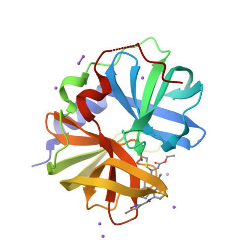
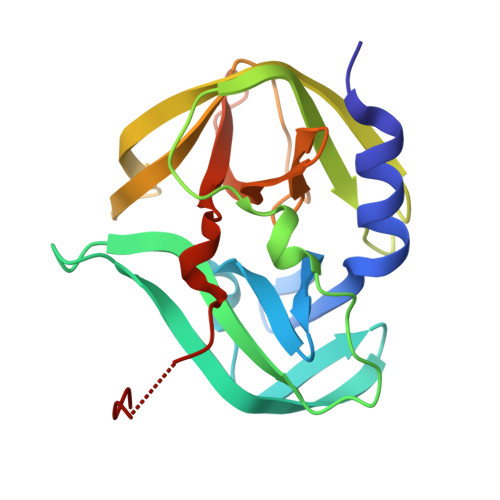
Structural Analysis of Inhibitor Binding to Enterovirus-D68 3C Protease.

Azzolino, V.N.Shaqra, A.M.Ali, A.Kurt Yilmaz, N.Schiffer, C.A.

(2025) Viruses 17

  • DOI: https://doi.org/10.3390/v17010075
  • Primary Citation of Related Structures:  
    8W3C, 8W3M, 8W3T

  • PubMed Abstract: 

    Enterovirus-D68 (EV68) continues to present as a global health issue causing respiratory illness and outbreaks associated with long-lasting neurological disease, with no antivirals or specific treatment options. The development of antiviral therapeutics, such as small-molecule inhibitors that target conserved proteins like the enteroviral 3C protease, remains to be achieved. While various 3C inhibitors have been investigated, their design does not consider the potential emergence of drug resistance mutations. For other antivirals where resistance has been a challenge, we have demonstrated that the likelihood of resistance can be minimized by designing inhibitors that leverage the evolutionary constraints of the target. Here, we characterize a series of 3C inhibitors against EV68-3C protease through enzyme inhibition, protein crystallography, and structural analysis. We have determined and analyzed three high-resolution inhibitor-bound crystal structures of EV68-3C protease, which revealed possible sites of resistance mutations, a key structural water molecule conserved during ligand binding, and the conformational flexibility of the catalytic histidine H40. This structural analysis combined with enzymatic assays provides insights for the rational design of inhibitors that are robust against resistance toward developing antiviral treatments for EV68 infections.


  • Organizational Affiliation

    Department of Biochemistry and Molecular Biotechnology, University of Massachusetts Chan Medical School, Worcester, MA 01605, USA.


Macromolecules
Find similar proteins by:  (by identity cutoff)  |  3D Structure
Entity ID: 1
MoleculeChains Sequence LengthOrganismDetailsImage
Peptidase C3190enterovirus D68Mutation(s): 0 
EC: 3.4.22.29 (PDB Primary Data), 3.6.1.15 (PDB Primary Data), 3.4.22.28 (PDB Primary Data), 2.7.7.48 (PDB Primary Data)
UniProt
Find proteins for A1E4A3 (Human enterovirus D68)
Explore A1E4A3 
Go to UniProtKB:  A1E4A3
Entity Groups  
Sequence Clusters30% Identity50% Identity70% Identity90% Identity95% Identity100% Identity
UniProt GroupA1E4A3
Sequence Annotations
Expand
  • Reference Sequence
Small Molecules
Ligands 2 Unique
IDChains Name / Formula / InChI Key2D Diagram3D Interactions
XNV (Subject of Investigation/LOI)
Query on XNV

Download Ideal Coordinates CCD File 
B [auth A]ethyl (4R)-4-({(2S)-2-[3-{[(5-methyl-1,2-oxazol-3-yl)carbonyl]amino}-2-oxopyridin-1(2H)-yl]pent-4-ynoyl}amino)-5-[(3S)-2-oxopyrrolidin-3-yl]pentanoate
C26 H31 N5 O7
WVFVHUYBPRBKRA-UEXGIBASSA-N
NA
Query on NA

Download Ideal Coordinates CCD File 
C [auth A]
D [auth A]
E [auth A]
F [auth A]
G [auth A]
SODIUM ION
Na
FKNQFGJONOIPTF-UHFFFAOYSA-N
Experimental Data & Validation

Experimental Data

  • Method: X-RAY DIFFRACTION
  • Resolution: 1.97 Å
  • R-Value Free:  0.238 (Depositor), 0.240 (DCC) 
  • R-Value Work:  0.206 (Depositor), 0.206 (DCC) 
  • R-Value Observed: 0.207 (Depositor) 
Space Group: P 31 2 1
Unit Cell:
Length ( Å )Angle ( ˚ )
a = 56.491α = 90
b = 56.491β = 90
c = 170.72γ = 120
Software Package:
Software NamePurpose
PHENIXrefinement
PHENIXphasing
Cootmodel building
XDSdata reduction
XDSdata scaling

Structure Validation

View Full Validation Report


Ligand Structure Quality Assessment 

Created with Raphaël 2.3.0Worse 01 BetterLigand structure goodness of fit to experimental dataBest fitted XNVClick on this verticalbar to view details

Entry History & Funding Information

Deposition Data


Funding OrganizationLocationGrant Number
National Institutes of Health/National Institute Of Allergy and Infectious Diseases (NIH/NIAID)United States1F30AI181515-01

Revision History  (Full details and data files)

  • Version 1.0: 2025-01-15
    Type: Initial release
  • Version 1.1: 2025-02-05
    Changes: Database references