8ZVH

Crystal structure of AetD in complex with L-phenylalanine


Experimental Data Snapshot

  • Method: X-RAY DIFFRACTION
  • Resolution: 1.87 Å
  • R-Value Free: 0.250 
  • R-Value Work: 0.192 
  • R-Value Observed: 0.195 

Starting Model: in silico
View more details

wwPDB Validation   3D Report Full Report


Ligand Structure Quality Assessment 


This is version 1.0 of the entry. See complete history


Literature

Structural and molecular insights of two unique enzymes involved in the biosynthesis of a natural halogenated nitrile.

Chen, C.C.Li, H.Huang, J.W.Guo, R.T.

(2024) FEBS J 

  • DOI: https://doi.org/10.1111/febs.17279
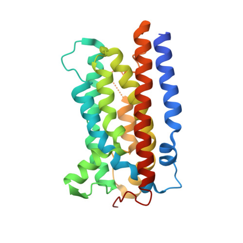
  • Primary Citation of Related Structures:  
    8ZVG, 8ZVH

  • PubMed Abstract: 

    Organohalogen compounds exhibit wide-ranging bioactivities and potential applications. Understanding natural biosynthetic pathways and improving the production of halogenated compounds has garnered significant attention. Recently, the biosynthetic pathway of a cyanobacterial neurotoxin, aetokthonotoxin, was reported. It contains two unique enzymes: a single-component flavin-dependent halogenase AetF and a new type of nitril synthase AetD. The crystal structures of these enzymes in complex with their cofactors and substrates that were recently reported will be presented here. The AetF structures reveal a tri-domain architecture, the transfer direction of the hydride ion, a possible path to deliver the hypohalous acid, and the unusual bispecific substrate-recognition mode. The AetD structures demonstrate that the nitrile formation should occur through the action of a diiron cluster, implying that the enzyme should be capable of catalyzing the nitrile formation of alternative amino acids. This information is of central importance for understanding the mechanism of action as well as the applications of these two the-first-of-its-kind enzymes.


  • Organizational Affiliation

    Zhejiang Key Laboratory of Medical Epigenetics, Department of Immunology and Pathogen Biology, School of Basic Medical Sciences, Hangzhou Normal University, Hangzhou, China.


Macromolecules
Find similar proteins by:  (by identity cutoff)  |  3D Structure
Entity ID: 1
MoleculeChains Sequence LengthOrganismDetailsImage
AetD
A, B
250Aetokthonos hydrillicola Thurmond2011Mutation(s): 0 
Gene Names: aetD
UniProt
Find proteins for A0A861B387 (Aetokthonos hydrillicola Thurmond2011)
Explore A0A861B387 
Go to UniProtKB:  A0A861B387
Entity Groups  
Sequence Clusters30% Identity50% Identity70% Identity90% Identity95% Identity100% Identity
UniProt GroupA0A861B387
Sequence Annotations
Expand
  • Reference Sequence
Small Molecules
Ligands 3 Unique
IDChains Name / Formula / InChI Key2D Diagram3D Interactions
PHE (Subject of Investigation/LOI)
Query on PHE

Download Ideal Coordinates CCD File 
D [auth A],
I [auth B]
PHENYLALANINE
C9 H11 N O2
COLNVLDHVKWLRT-QMMMGPOBSA-N
NI (Subject of Investigation/LOI)
Query on NI

Download Ideal Coordinates CCD File 
E [auth A],
F [auth A],
G [auth A],
J [auth B],
K [auth B]
NICKEL (II) ION
Ni
VEQPNABPJHWNSG-UHFFFAOYSA-N
FE2 (Subject of Investigation/LOI)
Query on FE2

Download Ideal Coordinates CCD File 
C [auth A],
H [auth B]
FE (II) ION
Fe
CWYNVVGOOAEACU-UHFFFAOYSA-N
Experimental Data & Validation

Experimental Data

  • Method: X-RAY DIFFRACTION
  • Resolution: 1.87 Å
  • R-Value Free: 0.250 
  • R-Value Work: 0.192 
  • R-Value Observed: 0.195 
  • Space Group: P 41
Unit Cell:
Length ( Å )Angle ( ˚ )
a = 65.043α = 90
b = 65.043β = 90
c = 116.211γ = 90
Software Package:
Software NamePurpose
REFMACrefinement
SAINTdata scaling
SAINTdata reduction
PHASERphasing

Structure Validation

View Full Validation Report


Ligand Structure Quality Assessment 


Entry History & Funding Information

Deposition Data


Funding OrganizationLocationGrant Number
National Natural Science Foundation of China (NSFC)China--

Revision History  (Full details and data files)

  • Version 1.0: 2024-10-02
    Type: Initial release