9AVS

Human alpha-galactosidase A in complex with saposin B


Experimental Data Snapshot

  • Method: X-RAY DIFFRACTION
  • Resolution: 3.53 Å
  • R-Value Free: 
    0.265 (Depositor), 0.267 (DCC) 
  • R-Value Work: 
    0.241 (Depositor), 0.241 (DCC) 
  • R-Value Observed: 
    0.242 (Depositor) 

Starting Model: experimental
View more details

wwPDB Validation   3D Report Full Report


Ligand Structure Quality Assessment 

Created with Raphaël 2.3.0Worse 01 BetterLigand structure goodness of fit to experimental dataBest fitted NAGClick on this verticalbar to view details

This is version 1.4 of the entry. See complete history


Literature

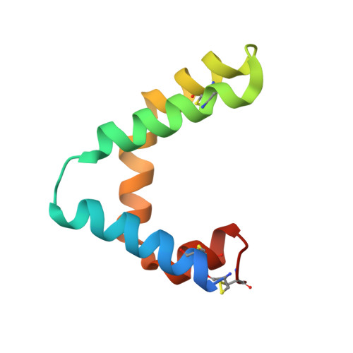
Human Saposin B Ligand Binding and Presentation to alpha-Galactosidase A.

Sawyer, T.K.Aral, E.Staros, J.V.Bobst, C.E.Garman, S.C.

(2024) bioRxiv 

  • DOI: https://doi.org/10.1101/2024.04.04.584535
  • Primary Citation of Related Structures:  
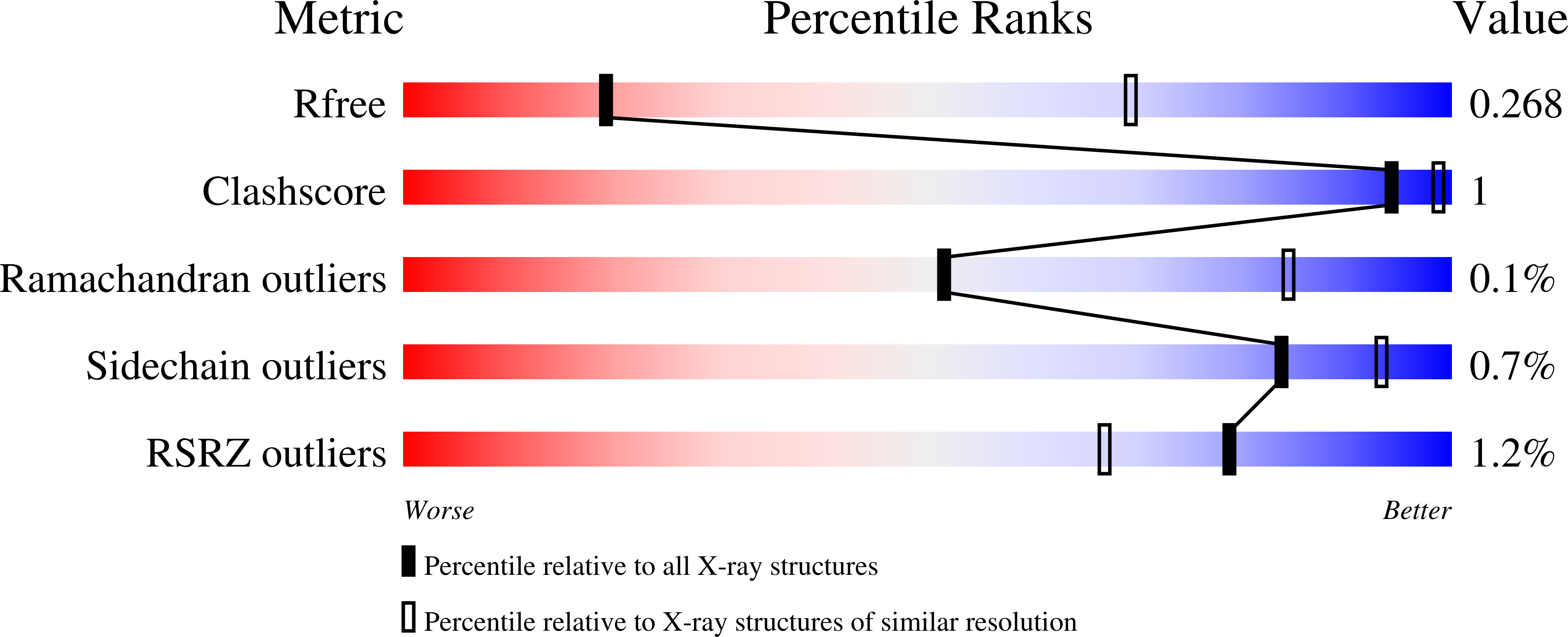
    9AVS, 9AXG

  • PubMed Abstract: 

    Sphingolipid activator protein B (saposin B; SapB) is an essential activator of globotriaosylceramide (Gb3) catabolism by α-galactosidase A. However, the manner by which SapB stimulates α-galactosidase A activity remains unknown. To uncover the molecular mechanism of SapB presenting Gb3 to α-galactosidase A, we subjected the fluorescent substrate globotriaosylceramide-nitrobenzoxidazole (Gb3-NBD) to a series of biochemical and structural assays involving SapB. First, we showed that SapB stably binds Gb3-NBD using a fluorescence equilibrium binding assay, isolates Gb3-NBD from micelles, and facilitates α-galactosidase A cleavage of Gb3-NBD in vitro . Second, we crystallized SapB in the presence of Gb3-NBD and validated the ligand-bound assembly. Third, we captured transient interactions between SapB and α-galactosidase A by chemical cross-linking. Finally, we determined the crystal structure of SapB bound to α-galactosidase A. These findings establish general principles for molecular recognition in saposin:hydrolase complexes and highlight the utility of NBD reporter lipids in saposin biochemistry and structural biology.


  • Organizational Affiliation

    Department of Biochemistry & Molecular Biology, Institute of Applied Life Sciences, University of Massachusetts, Amherst, Massachusetts 01003, USA.


Macromolecules
Find similar proteins by:  (by identity cutoff)  |  3D Structure
Entity ID: 1
MoleculeChains Sequence LengthOrganismDetailsImage
Alpha-galactosidase A
A, B
414Homo sapiensMutation(s): 1 
Gene Names: GLA
EC: 3.2.1.22
UniProt & NIH Common Fund Data Resources
Find proteins for P06280 (Homo sapiens)
Explore P06280 
Go to UniProtKB:  P06280
PHAROS:  P06280
GTEx:  ENSG00000102393 
Entity Groups  
Sequence Clusters30% Identity50% Identity70% Identity90% Identity95% Identity100% Identity
UniProt GroupP06280
Glycosylation
Glycosylation Sites: 3Go to GlyGen: P06280-1
Sequence Annotations
Expand
  • Reference Sequence
Find similar proteins by:  (by identity cutoff)  |  3D Structure
Entity ID: 2
MoleculeChains Sequence LengthOrganismDetailsImage
Saposin-B82Homo sapiensMutation(s): 0 
Gene Names: PSAPGLBASAP1
UniProt & NIH Common Fund Data Resources
Find proteins for P07602 (Homo sapiens)
Explore P07602 
Go to UniProtKB:  P07602
PHAROS:  P07602
GTEx:  ENSG00000197746 
Entity Groups  
Sequence Clusters30% Identity50% Identity70% Identity90% Identity95% Identity100% Identity
UniProt GroupP07602
Sequence Annotations
Expand
  • Reference Sequence
Oligosaccharides

Help

Entity ID: 3
MoleculeChains Length2D Diagram Glycosylation3D Interactions
alpha-D-mannopyranose-(1-3)-beta-D-mannopyranose-(1-4)-2-acetamido-2-deoxy-beta-D-glucopyranose-(1-4)-[alpha-L-fucopyranose-(1-6)]2-acetamido-2-deoxy-beta-D-glucopyranoseD [auth E]5N-Glycosylation
Glycosylation Resources
GlyTouCan:  G42466VF
GlyCosmos:  G42466VF
GlyGen:  G42466VF
Entity ID: 4
MoleculeChains Length2D Diagram Glycosylation3D Interactions
alpha-D-mannopyranose-(1-3)-beta-D-mannopyranose-(1-4)-2-acetamido-2-deoxy-beta-D-glucopyranose-(1-4)-2-acetamido-2-deoxy-beta-D-glucopyranoseE [auth D],
G
4N-Glycosylation
Glycosylation Resources
GlyTouCan:  G81315DD
GlyCosmos:  G81315DD
GlyGen:  G81315DD
Entity ID: 5
MoleculeChains Length2D Diagram Glycosylation3D Interactions
2-acetamido-2-deoxy-beta-D-glucopyranose-(1-4)-2-acetamido-2-deoxy-beta-D-glucopyranose
F
2N-Glycosylation
Glycosylation Resources
GlyTouCan:  G42666HT
GlyCosmos:  G42666HT
GlyGen:  G42666HT
Entity ID: 6
MoleculeChains Length2D Diagram Glycosylation3D Interactions
alpha-D-mannopyranose-(1-3)-[alpha-D-mannopyranose-(1-6)]beta-D-mannopyranose-(1-4)-2-acetamido-2-deoxy-beta-D-glucopyranose-(1-4)-2-acetamido-2-deoxy-beta-D-glucopyranose
H
5N-Glycosylation
Glycosylation Resources
GlyTouCan:  G22768VO
GlyCosmos:  G22768VO
GlyGen:  G22768VO
Small Molecules
Ligands 2 Unique
IDChains Name / Formula / InChI Key2D Diagram3D Interactions
NAG
Query on NAG

Download Ideal Coordinates CCD File 
Q [auth B]2-acetamido-2-deoxy-beta-D-glucopyranose
C8 H15 N O6
OVRNDRQMDRJTHS-FMDGEEDCSA-N
SO4
Query on SO4

Download Ideal Coordinates CCD File 
I [auth A]
J [auth A]
K [auth A]
L [auth A]
M [auth A]
SULFATE ION
O4 S
QAOWNCQODCNURD-UHFFFAOYSA-L
Experimental Data & Validation

Experimental Data

  • Method: X-RAY DIFFRACTION
  • Resolution: 3.53 Å
  • R-Value Free:  0.265 (Depositor), 0.267 (DCC) 
  • R-Value Work:  0.241 (Depositor), 0.241 (DCC) 
  • R-Value Observed: 0.242 (Depositor) 
Space Group: C 2 2 21
Unit Cell:
Length ( Å )Angle ( ˚ )
a = 120.116α = 90
b = 142.563β = 90
c = 191.967γ = 90
Software Package:
Software NamePurpose
PHASERphasing
PHENIXrefinement
xia2data reduction
xia2data scaling

Structure Validation

View Full Validation Report


Ligand Structure Quality Assessment 

Created with Raphaël 2.3.0Worse 01 BetterLigand structure goodness of fit to experimental dataBest fitted NAGClick on this verticalbar to view details

Entry History & Funding Information

Deposition Data


Funding OrganizationLocationGrant Number
National Institutes of Health/National Institute of Diabetes and Digestive and Kidney Disease (NIH/NIDDK)United States5R01DK076877

Revision History  (Full details and data files)

  • Version 1.0: 2024-04-17
    Type: Initial release
  • Version 1.1: 2024-04-24
    Changes: Database references
  • Version 1.2: 2024-08-21
    Changes: Experimental preparation
  • Version 1.3: 2024-09-04
    Changes: Database references
  • Version 1.4: 2024-10-23
    Changes: Structure summary