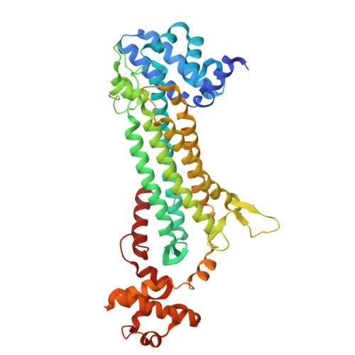
9GGJ

Crystal structure of argininosuccinate lyase from Arabidopsis thaliana (AtASL) in complex with biological substrate and products - argininosuccinate, argnine and fumarate


Experimental Data Snapshot

  • Method: X-RAY DIFFRACTION
  • Resolution: 2.00 Å
  • R-Value Free: 0.227 
  • R-Value Work: 0.175 
  • R-Value Observed: 0.176 

Starting Model: experimental
View more details

wwPDB Validation   3D Report Full Report


Ligand Structure Quality Assessment 


This is version 1.1 of the entry. See complete history


Literature

Arabidopsis thaliana argininosuccinate lyase structure uncovers the role of serine as the catalytic base.

Nielipinski, M.Nielipinska, D.Pietrzyk-Brzezinska, A.J.Sekula, B.

(2024) J Struct Biol 216: 108130-108130

  • DOI: https://doi.org/10.1016/j.jsb.2024.108130
  • Primary Citation of Related Structures:  
    9GGI, 9GGJ

  • PubMed Abstract: 

    Arginine is an important amino acid in plants, as it not only plays a structural role and serves as nitrogen storage but is also a precursor for various molecules, including polyamines and proline. Arginine is produced by argininosuccinate lyase (ASL) which catalyzes the cleavage of argininosuccinate to arginine and fumarate. ASL belongs to the fumarate lyase family and while many members of this family were well-characterized, little is known about plant ASLs. Here we present the first crystal structures of ASL from the model plant, Arabidopsis thaliana (AtASL). One of the structures represents the unliganded form of the AtASL homotetramer. The other structure, obtained from a crystal soaked in argininosuccinate, accommodates the substrate or the reaction products in one of four active sites of the AtASL tetramer. Each active site is located at the interface of three neighboring protomers. The AtASL structure with ligands allowed us to analyze the enzyme-substrate and the enzyme-product interactions in detail. Furthermore, based on our analyses, we describe residues of AtASL crucial for catalysis. The structure of AtASL gives the rationale for the open-to-close transition of the GSS mobile loop and indicates the importance of serine 333 from this loop for the enzymatic action of the enzyme. Finally, we supplemented the structural data with the identification of sequence motifs characteristic for ASLs.


  • Organizational Affiliation

    Institute of Molecular and Industrial Biotechnology, Faculty of Biotechnology and Food Sciences, Lodz University of Technology, Poland.


Macromolecules
Find similar proteins by:  (by identity cutoff)  |  3D Structure
Entity ID: 1
MoleculeChains Sequence LengthOrganismDetailsImage
Argininosuccinate lyase, chloroplastic
A, B, C, D, E
A, B, C, D, E, F, G, H
465Arabidopsis thalianaMutation(s): 0 
Gene Names: At5g10920T30N20.190
EC: 4.3.2.1
UniProt
Find proteins for Q9LEU8 (Arabidopsis thaliana)
Explore Q9LEU8 
Go to UniProtKB:  Q9LEU8
Entity Groups  
Sequence Clusters30% Identity50% Identity70% Identity90% Identity95% Identity100% Identity
UniProt GroupQ9LEU8
Sequence Annotations
Expand
  • Reference Sequence
Small Molecules
Ligands 6 Unique
IDChains Name / Formula / InChI Key2D Diagram3D Interactions
AS1 (Subject of Investigation/LOI)
Query on AS1

Download Ideal Coordinates CCD File 
DA [auth D],
HA [auth E],
I [auth A],
TA [auth G],
YA [auth H]
ARGININOSUCCINATE
C10 H18 N4 O6
KDZOASGQNOPSCU-WDSKDSINSA-N
ARG
Query on ARG

Download Ideal Coordinates CCD File 
CA [auth C],
SA [auth F],
V [auth B]
ARGININE
C6 H15 N4 O2
ODKSFYDXXFIFQN-BYPYZUCNSA-O
FUM (Subject of Investigation/LOI)
Query on FUM

Download Ideal Coordinates CCD File 
IA [auth E],
Q [auth A]
FUMARIC ACID
C4 H4 O4
VZCYOOQTPOCHFL-OWOJBTEDSA-N
SO4
Query on SO4

Download Ideal Coordinates CCD File 
AB [auth H]
BB [auth H]
CB [auth H]
FA [auth D]
GA [auth D]
AB [auth H],
BB [auth H],
CB [auth H],
FA [auth D],
GA [auth D],
J [auth A],
JA [auth E],
K [auth A],
KA [auth E],
L [auth A],
LA [auth E],
M [auth A],
MA [auth E],
N [auth A],
OA [auth F],
PA [auth F],
QA [auth F],
R [auth B],
RA [auth F],
S [auth B],
T [auth B],
U [auth B],
W [auth C],
WA [auth G],
X [auth C],
Y [auth C],
ZA [auth H]
SULFATE ION
O4 S
QAOWNCQODCNURD-UHFFFAOYSA-L
GOL
Query on GOL

Download Ideal Coordinates CCD File 
EA [auth D],
UA [auth G],
VA [auth G]
GLYCEROL
C3 H8 O3
PEDCQBHIVMGVHV-UHFFFAOYSA-N
CL
Query on CL

Download Ideal Coordinates CCD File 
AA [auth C]
BA [auth C]
NA [auth E]
O [auth A]
P [auth A]
AA [auth C],
BA [auth C],
NA [auth E],
O [auth A],
P [auth A],
XA [auth G],
Z [auth C]
CHLORIDE ION
Cl
VEXZGXHMUGYJMC-UHFFFAOYSA-M
Experimental Data & Validation

Experimental Data

  • Method: X-RAY DIFFRACTION
  • Resolution: 2.00 Å
  • R-Value Free: 0.227 
  • R-Value Work: 0.175 
  • R-Value Observed: 0.176 
  • Space Group: P 1 21 1
Unit Cell:
Length ( Å )Angle ( ˚ )
a = 106.588α = 90
b = 230.848β = 89.48
c = 112.534γ = 90
Software Package:
Software NamePurpose
REFMACrefinement
XDSdata reduction
STARANISOdata scaling
PHASERphasing

Structure Validation

View Full Validation Report


Ligand Structure Quality Assessment 


Entry History & Funding Information

Deposition Data


Funding OrganizationLocationGrant Number
Polish National Science CentrePolandSONATA17 2021/43/D/NZ1/00486

Revision History  (Full details and data files)

  • Version 1.0: 2024-10-23
    Type: Initial release
  • Version 1.1: 2024-10-30
    Changes: Database references