


Identification and Characterization of Pyrimidine Nucleoside 2'-Hydroxylase
Genz, F., Friedrich, F., Lonarz, C., Einsle, O., Jung, M., Muller, M., Fessner, N.D.(2025) ACS Catal
Experimental Data Snapshot
Starting Model: in silico
View more details
(2025) ACS Catal
Entity ID: 1 | |||||
|---|---|---|---|---|---|
| Molecule | Chains | Sequence Length | Organism | Details | Image |
| Clavaminate synthase-like protein | 390 | Neurospora crassa | Mutation(s): 0 Gene Names: NCU02560 | 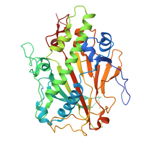 | |
UniProt | |||||
Find proteins for Q7SHQ5 (Neurospora crassa (strain ATCC 24698 / 74-OR23-1A / CBS 708.71 / DSM 1257 / FGSC 987)) Explore Q7SHQ5 Go to UniProtKB: Q7SHQ5 | |||||
Entity Groups | |||||
| Sequence Clusters | 30% Identity50% Identity70% Identity90% Identity95% Identity100% Identity | ||||
| UniProt Group | Q7SHQ5 | ||||
Sequence AnnotationsExpand | |||||
| |||||
| Ligands 5 Unique | |||||
|---|---|---|---|---|---|
| ID | Chains | Name / Formula / InChI Key | 2D Diagram | 3D Interactions | |
| THM (Subject of Investigation/LOI) Query on THM | D [auth A], H [auth B] | THYMIDINE C10 H14 N2 O5 IQFYYKKMVGJFEH-XLPZGREQSA-N |  | ||
| BU3 Query on BU3 | I [auth B] | (R,R)-2,3-BUTANEDIOL C4 H10 O2 OWBTYPJTUOEWEK-QWWZWVQMSA-N |  | ||
| EDO Query on EDO | E [auth A] | 1,2-ETHANEDIOL C2 H6 O2 LYCAIKOWRPUZTN-UHFFFAOYSA-N |  | ||
| MN Query on MN | C [auth A], F [auth B] | MANGANESE (II) ION Mn WAEMQWOKJMHJLA-UHFFFAOYSA-N |  | ||
| CL Query on CL | G [auth B] | CHLORIDE ION Cl VEXZGXHMUGYJMC-UHFFFAOYSA-M |  | ||
| Length ( Å ) | Angle ( ˚ ) |
|---|---|
| a = 57.983 | α = 90 |
| b = 108.293 | β = 90 |
| c = 115.732 | γ = 90 |
| Software Name | Purpose |
|---|---|
| PHENIX | refinement |
| Aimless | data scaling |
| autoPROC | data reduction |
| PHASER | phasing |
| Funding Organization | Location | Grant Number |
|---|---|---|
| Not funded | -- |
RCSB PDB is hosted by

RCSB PDB is a member of the


