9IT8 | pdb_00009it8

Crystal structure of the ternary complex of lactoperoxidase with nitric oxide and nitrite ion at 1.95 A resolution


Experimental Data Snapshot

  • Method: X-RAY DIFFRACTION
  • Resolution: 1.95 Å
  • R-Value Free: 
    0.247 (Depositor), 0.252 (DCC) 
  • R-Value Work: 
    0.188 (Depositor), 0.198 (DCC) 

Starting Model: experimental
View more details

wwPDB Validation   3D Report Full Report


Ligand Structure Quality Assessment 

Created with Raphaël 2.3.0Worse 01 BetterLigand structure goodness of fit to experimental dataBest fitted HEMClick on this verticalbar to view detailsBest fitted NAGClick on this verticalbar to view detailsBest fitted EDOClick on this verticalbar to view details

This is version 1.2 of the entry. See complete history


Literature

Structure of the Complex of Lactoperoxidase With Nitric Oxide at 1.95 angstrom Resolution.

Maurya, A.Ahmad, N.Sharma, P.Sharma, S.Singh, T.P.

(2025) Proteins 

  • DOI: https://doi.org/10.1002/prot.26797
  • Primary Citation of Related Structures:  
    9IT8

  • PubMed Abstract: 

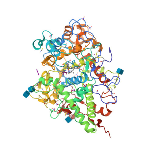
    Lactoperoxidase (LPO) is a heme-containing mammalian enzyme that is found in the extracellular fluids of animals including plasma, saliva, airway epithelial and nasal lining fluids, milk, tears, and gastric juices. LPO uses hydrogen peroxide (H 2 O 2 ) to convert substrates into oxidized products. Previous structural studies have shown that H 2 O 2 , CO, and CN are bound to LPO at the distal heme cavity by coordinating with heme iron. The structure of the complex of LPO with NO shows that NO also binds to LPO at the distal heme cavity and forms a coordinate linkage with heme iron. The structure shows that the nitrogen atom of NO is linked to heme iron at a distance of 1.97 while the oxygen atom is attached to the N ε2 atom of His109 at a distance of 2.23 Å. On the other hand, N atom of NO is located with an interatomic distance of 3.25 Å allowing a hydrogen-bonding interaction with the N ε2 atom of Gln105. A comparison of the bindings of NO, CO, CN, and H 2 O 2 in coordination with heme iron indicates stereochemical compatibility of the distal heme cavity for the binding of diatomic molecules. However, notable differences are observed in their orientations in the distal heme cavity indicating functional differences. The bindings of NO, CO, and CN by coordinating with heme iron result in the inhibition of LPO while the binding of H 2 O 2 to heme iron produces an intermediate of LPO known as Compound I.


  • Organizational Affiliation

    Department of Biophysics, All India Institute of Medical Sciences, New Delhi, India.


Macromolecules
Find similar proteins by:  (by identity cutoff)  |  3D Structure
Entity ID: 1
MoleculeChains Sequence LengthOrganismDetailsImage
Lactoperoxidase595Bos taurusMutation(s): 0 
EC: 1.11.1.7
UniProt
Find proteins for P80025 (Bos taurus)
Explore P80025 
Go to UniProtKB:  P80025
Entity Groups  
Sequence Clusters30% Identity50% Identity70% Identity90% Identity95% Identity100% Identity
UniProt GroupP80025
Glycosylation
Glycosylation Sites: 4
Sequence Annotations
Expand
  • Reference Sequence
Oligosaccharides

Help

Entity ID: 2
MoleculeChains Length2D Diagram Glycosylation3D Interactions
2-acetamido-2-deoxy-beta-D-glucopyranose-(1-4)-2-acetamido-2-deoxy-beta-D-glucopyranose
B
2N-Glycosylation
Glycosylation Resources
GlyTouCan:  G42666HT
GlyCosmos:  G42666HT
GlyGen:  G42666HT
Small Molecules
Ligands 10 Unique
IDChains Name / Formula / InChI Key2D Diagram3D Interactions
HEM (Subject of Investigation/LOI)
Query on HEM

Download Ideal Coordinates CCD File 
Z [auth A]PROTOPORPHYRIN IX CONTAINING FE
C34 H32 Fe N4 O4
KABFMIBPWCXCRK-RGGAHWMASA-L
NAG (Subject of Investigation/LOI)
Query on NAG

Download Ideal Coordinates CCD File 
C [auth A],
D [auth A],
E [auth A]
2-acetamido-2-deoxy-beta-D-glucopyranose
C8 H15 N O6
OVRNDRQMDRJTHS-FMDGEEDCSA-N
IOD (Subject of Investigation/LOI)
Query on IOD

Download Ideal Coordinates CCD File 
EA [auth A]
FA [auth A]
GA [auth A]
HA [auth A]
NA [auth A]
IODIDE ION
I
XMBWDFGMSWQBCA-UHFFFAOYSA-M
OSM (Subject of Investigation/LOI)
Query on OSM

Download Ideal Coordinates CCD File 
DA [auth A]1-(OXIDOSULFANYL)METHANAMINE
C H5 N O S
KONFWJXIYYGXOO-UHFFFAOYSA-N
EDO (Subject of Investigation/LOI)
Query on EDO

Download Ideal Coordinates CCD File 
AA [auth A]1,2-ETHANEDIOL
C2 H6 O2
LYCAIKOWRPUZTN-UHFFFAOYSA-N
NO3 (Subject of Investigation/LOI)
Query on NO3

Download Ideal Coordinates CCD File 
H [auth A]
I [auth A]
IA [auth A]
M [auth A]
N [auth A]
NITRATE ION
N O3
NHNBFGGVMKEFGY-UHFFFAOYSA-N
SCN (Subject of Investigation/LOI)
Query on SCN

Download Ideal Coordinates CCD File 
CA [auth A]
J [auth A]
K [auth A]
L [auth A]
LA [auth A]
THIOCYANATE ION
C N S
ZMZDMBWJUHKJPS-UHFFFAOYSA-M
NO2 (Subject of Investigation/LOI)
Query on NO2

Download Ideal Coordinates CCD File 
G [auth A],
KA [auth A]
NITRITE ION
N O2
IOVCWXUNBOPUCH-UHFFFAOYSA-M
CA (Subject of Investigation/LOI)
Query on CA

Download Ideal Coordinates CCD File 
BA [auth A]CALCIUM ION
Ca
BHPQYMZQTOCNFJ-UHFFFAOYSA-N
NO (Subject of Investigation/LOI)
Query on NO

Download Ideal Coordinates CCD File 
F [auth A],
JA [auth A]
NITRIC OXIDE
N O
ODUCDPQEXGNKDN-UHFFFAOYSA-N
Experimental Data & Validation

Experimental Data

  • Method: X-RAY DIFFRACTION
  • Resolution: 1.95 Å
  • R-Value Free:  0.247 (Depositor), 0.252 (DCC) 
  • R-Value Work:  0.188 (Depositor), 0.198 (DCC) 
Space Group: P 1 21 1
Unit Cell:
Length ( Å )Angle ( ˚ )
a = 54.048α = 90
b = 80.503β = 102.849
c = 75.951γ = 90
Software Package:
Software NamePurpose
REFMACrefinement
XDSdata reduction
Aimlessdata scaling
MOLREPphasing

Structure Validation

View Full Validation Report


Ligand Structure Quality Assessment 

Created with Raphaël 2.3.0Worse 01 BetterLigand structure goodness of fit to experimental dataBest fitted HEMClick on this verticalbar to view detailsBest fitted NAGClick on this verticalbar to view detailsBest fitted EDOClick on this verticalbar to view details

Entry History & Funding Information

Deposition Data


Funding OrganizationLocationGrant Number
Indian Council of Medical ResearchIndiaIR-661

Revision History  (Full details and data files)

  • Version 1.0: 2024-09-11
    Type: Initial release
  • Version 1.1: 2024-10-09
    Changes: Structure summary
  • Version 1.2: 2025-03-19
    Changes: Database references