1G97 | pdb_00001g97

S.PNEUMONIAE GLMU COMPLEXED WITH UDP-N-ACETYLGLUCOSAMINE AND MG2+


Experimental Data Snapshot

  • Method: X-RAY DIFFRACTION
  • Resolution: 1.96 Å
  • R-Value Free: 
    0.221 (Depositor) 
  • R-Value Work: 
    0.188 (Depositor), 0.190 (DCC) 

Starting Model: experimental
View more details

wwPDB Validation   3D Report Full Report


Ligand Structure Quality Assessment 

Created with Raphaël 2.3.0Worse 01 BetterLigand structure goodness of fit to experimental dataBest fitted UD1Click on this verticalbar to view details

This is version 1.3 of the entry. See complete history


Literature

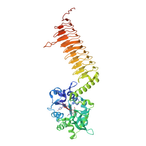
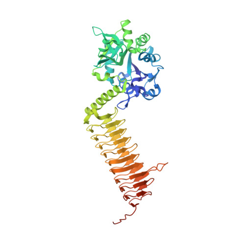
Crystal structures of Streptococcus pneumoniae N-acetylglucosamine-1-phosphate uridyltransferase, GlmU, in apo form at 2.33 A resolution and in complex with UDP-N-acetylglucosamine and Mg(2+) at 1.96 A resolution.

Kostrewa, D.D'Arcy, A.Takacs, B.Kamber, M.

(2001) J Mol Biology 305: 279-289

  • DOI: https://doi.org/10.1006/jmbi.2000.4296
  • Primary Citation of Related Structures:  
    1G95, 1G97

  • PubMed Abstract: 

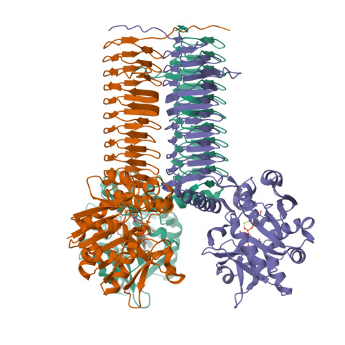
    N-Acetylglucosamine-1-phosphate uridyltransferase (GlmU) is an essential bacterial enzyme with both an acetyltransferase and a uridyltransferase activity which have been mapped to the C-terminal and N-terminal domains, respectively. GlmU performs the last two steps in the synthesis of UDP-N-acetylglucosamine (UDP-GlcNAc), which is an essential precursor in both the peptidoglycan and the lipopolysaccharide metabolic pathways. GlmU is therefore an attractive target for potential antibiotics. Knowledge of its three-dimensional structure would provide a basis for rational drug design. We have determined the crystal structures of Streptococcus pneumoniae GlmU (SpGlmU) in apo form at 2.33 A resolution, and in complex with UDP-N-acetyl glucosamine and the essential co-factor Mg(2+) at 1.96 A resolution. The protein structure consists of an N-terminal domain with an alpha/beta-fold, containing the uridyltransferase active site, and a C-terminal domain with a long left-handed beta-sheet helix (LbetaH) domain. An insertion loop containing the highly conserved sequence motif Asn-Tyr-Asp-Gly protrudes from the left-handed beta-sheet helix domain. In the crystal, S. pneumoniae GlmU forms exact trimers, mainly through contacts between left-handed beta-sheet helix domains. UDP-N-acetylglucosamine and Mg(2+) are bound at the uridyltransferase active site, which is in a closed form. We propose a uridyltransferase mechanism in which the activation energy of the double negatively charged phosphorane transition state is lowered by charge compensation of Mg(2+) and the side-chain of Lys22.


  • Organizational Affiliation

    Pharmaceutical Research Chemical Technologies, F. Hoffmann-La Roche Ltd., Basle, 4070, Switzerland. dirk.kostrewa@psi.ch


Macromolecules
Find similar proteins by:  (by identity cutoff)  |  3D Structure
Entity ID: 1
MoleculeChains Sequence LengthOrganismDetailsImage
N-ACETYLGLUCOSAMINE-1-PHOSPHATE URIDYLTRANSFERASE459Streptococcus pneumoniaeMutation(s): 0 
EC: 2.7.7.23 (PDB Primary Data), 2.3.1.157 (UniProt)
UniProt
Find proteins for Q97R46 (Streptococcus pneumoniae serotype 4 (strain ATCC BAA-334 / TIGR4))
Explore Q97R46 
Go to UniProtKB:  Q97R46
Entity Groups  
Sequence Clusters30% Identity50% Identity70% Identity90% Identity95% Identity100% Identity
UniProt GroupQ97R46
Sequence Annotations
Expand
  • Reference Sequence
Experimental Data & Validation

Experimental Data

  • Method: X-RAY DIFFRACTION
  • Resolution: 1.96 Å
  • R-Value Free:  0.221 (Depositor) 
  • R-Value Work:  0.188 (Depositor), 0.190 (DCC) 
Space Group: H 3 2
Unit Cell:
Length ( Å )Angle ( ˚ )
a = 93.66α = 90
b = 93.66β = 90
c = 275.9γ = 120
Software Package:
Software NamePurpose
CNSrefinement
DENZOdata reduction
SCALEPACKdata scaling
CNSphasing

Structure Validation

View Full Validation Report


Ligand Structure Quality Assessment 

Created with Raphaël 2.3.0Worse 01 BetterLigand structure goodness of fit to experimental dataBest fitted UD1Click on this verticalbar to view details

Entry History 

Deposition Data

Revision History  (Full details and data files)

  • Version 1.0: 2001-05-22
    Type: Initial release
  • Version 1.1: 2008-04-27
    Changes: Version format compliance
  • Version 1.2: 2011-07-13
    Changes: Derived calculations, Version format compliance
  • Version 1.3: 2023-08-09
    Changes: Data collection, Database references, Derived calculations, Refinement description