1H0V | pdb_00001h0v

Human cyclin dependent protein kinase 2 in complex with the inhibitor 2-Amino-6-[(R)-pyrrolidino-5'-yl]methoxypurine


Experimental Data Snapshot

  • Method: X-RAY DIFFRACTION
  • Resolution: 1.90 Å
  • R-Value Free: 
    0.234 (Depositor) 
  • R-Value Work: 
    0.154 (Depositor), 0.170 (DCC) 
  • R-Value Observed: 
    0.158 (Depositor) 

Starting Model: experimental
View more details

wwPDB Validation   3D Report Full Report


Ligand Structure Quality Assessment 

Created with Raphaël 2.3.0Worse 01 BetterLigand structure goodness of fit to experimental dataBest fitted UN4Click on this verticalbar to view details

This is version 1.4 of the entry. See complete history


Literature

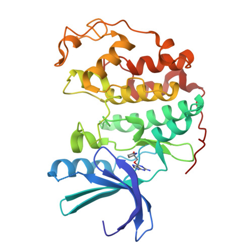
Probing the ATP Ribose-Binding Domain of Cyclin-Dependent Kinases 1 and 2 with O(6)-Substituted Guanine Derivatives

Gibson, A.E.Arris, C.E.Bentley, J.Boyle, F.T.Curtin, N.J.Davies, T.G.Endicott, J.A.Golding, B.T.Grant, S.Griffin, R.J.Jewsbury, P.Johnson, L.N.Mesguiche, V.Newell, D.R.Noble, M.E.M.Tucker, J.A.Whitfield, H.J.

(2002) J Med Chem 45: 3381

  • DOI: https://doi.org/10.1021/jm020056z
  • Primary Citation of Related Structures:  
    1GZ8, 1H0V, 1H0W

  • PubMed Abstract: 

    O(6)-substituted guanines are adenosine 5'-triphosphate (ATP) competitive inhibitors of CDK1/cyclin B1 and CDK2/cyclin A, the O(6) substituent occupying the kinase ribose binding site. Fifty-eight O(6)-substituted guanines were prepared to probe the ribose pocket, and the structures of four representative compounds bound to monomeric CDK2 were determined by X-ray crystallography. Optimum binding occurs with a moderately sized aliphatic O(6) substituent that packs tightly against the hydrophobic patch presented by the glycine loop, centered on Val18, an interaction promoted by the conformational restraints imposed in a cyclohexylmethyl or cyclohexenylmethyl ring. Structure-based design generated (R)-(2-amino-9H-purin-6-yloxymethyl)pyrrolidin-2-one (56), which reproduces the reported hydrogen bonds formed between ATP and Asp86 and Gln131 but failed to improve inhibitory potency. Thus, the parent compound O(6)-cyclohexylmethylguanine (NU2058, 25) is the preferred starting point for exploring other areas of the kinase active site.


  • Organizational Affiliation

    Department of Chemistry and Cancer Research Unit, University of Newcastle, Newcastle upon Tyne NE2 4HH, United Kingdom.


Macromolecules
Find similar proteins by:  (by identity cutoff)  |  3D Structure
Entity ID: 1
MoleculeChains Sequence LengthOrganismDetailsImage
CELL DIVISION PROTEIN KINASE 2298Homo sapiensMutation(s): 0 
EC: 2.7.1.37 (PDB Primary Data), 2.7.11.22 (UniProt)
UniProt & NIH Common Fund Data Resources
Find proteins for P24941 (Homo sapiens)
Explore P24941 
Go to UniProtKB:  P24941
PHAROS:  P24941
GTEx:  ENSG00000123374 
Entity Groups  
Sequence Clusters30% Identity50% Identity70% Identity90% Identity95% Identity100% Identity
UniProt GroupP24941
Sequence Annotations
Expand
  • Reference Sequence
Small Molecules
Ligands 1 Unique
IDChains Name / Formula / InChI Key2D Diagram3D Interactions
UN4
Query on UN4

Download Ideal Coordinates CCD File 
B [auth A]5-{[(2-AMINO-9H-PURIN-6-YL)OXY]METHYL}-2-PYRROLIDINONE
C10 H12 N6 O2
UYPMMHCTXQIWDX-RXMQYKEDSA-N
Experimental Data & Validation

Experimental Data

  • Method: X-RAY DIFFRACTION
  • Resolution: 1.90 Å
  • R-Value Free:  0.234 (Depositor) 
  • R-Value Work:  0.154 (Depositor), 0.170 (DCC) 
  • R-Value Observed: 0.158 (Depositor) 
Space Group: P 21 21 21
Unit Cell:
Length ( Å )Angle ( ˚ )
a = 52.953α = 90
b = 70.912β = 90
c = 72.283γ = 90
Software Package:
Software NamePurpose
REFMACrefinement
DENZOdata reduction
SCALEPACKdata scaling
CCP4phasing

Structure Validation

View Full Validation Report


Ligand Structure Quality Assessment 

Created with Raphaël 2.3.0Worse 01 BetterLigand structure goodness of fit to experimental dataBest fitted UN4Click on this verticalbar to view details

Entry History 

Revision History  (Full details and data files)

  • Version 1.0: 2003-06-27
    Type: Initial release
  • Version 1.1: 2011-05-08
    Changes: Version format compliance
  • Version 1.2: 2011-07-13
    Changes: Version format compliance
  • Version 1.3: 2019-03-06
    Changes: Data collection, Experimental preparation, Other
  • Version 1.4: 2023-12-13
    Changes: Data collection, Database references, Refinement description