1W7C | pdb_00001w7c

PPLO at 1.23 Angstroms


Experimental Data Snapshot

  • Method: X-RAY DIFFRACTION
  • Resolution: 1.23 Å
  • R-Value Free: 
    0.146 (Depositor), 0.160 (DCC) 
  • R-Value Work: 
    0.112 (Depositor), 0.130 (DCC) 
  • R-Value Observed: 
    0.113 (Depositor) 

Starting Model: experimental
View more details

wwPDB Validation   3D Report Full Report


Ligand Structure Quality Assessment 

Created with Raphaël 2.3.0Worse 01 BetterLigand structure goodness of fit to experimental dataBest fitted NAGClick on this verticalbar to view details

This is version 2.1 of the entry. See complete history


Literature

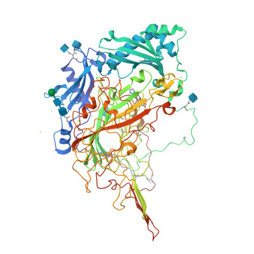
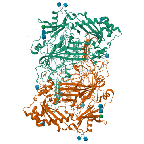
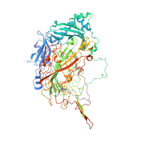
The 1.23 A Structure of Pichia Pastoris Lysyl Oxidase Reveals a Lysine-Lysine Cross-Link

Duff, A.P.Cohen, A.E.Ellis, P.J.Hilmer, K.Langley, D.B.Dooley, D.M.Freeman, H.C.Guss, J.M.

(2006) Acta Crystallogr D Biol Crystallogr 62: 1073

  • DOI: https://doi.org/10.1107/S0907444906026333
  • Primary Citation of Related Structures:  
    1W7C

  • PubMed Abstract: 

    The structure of Pichia pastoris lysyl oxidase (PPLO) in a new crystal form has been refined at 1.23 Angstrom resolution. PPLO, a copper amine oxidase (CuAO) with a 2,4,5-trihydroxyphenylalanine quinone (TPQ) cofactor, differs from most other members of the CuAO enzyme family in having the ability to oxidize the side chain of lysine residues in a polypeptide. In the asymmetric unit of the crystals, the structure analysis has located residues 43-779 of the polypeptide chain, seven carbohydrate residues, the active-site Cu atom, an imidazole molecule bound at the active site, two buried Ca(2+) ions, five surface Mg(2+) ions, five surface Cl(-) ions and 1045 water molecules. The crystallographic residuals are R = 0.112 and R(free) = 0.146. The TPQ cofactor and several other active-site residues are poorly ordered, in contrast to the surrounding well ordered structure. A covalent cross-link is observed between two lysine residues, Lys778 and Lys66. The cross-link is likely to have been formed by the oxidation of Lys778 followed by a spontaneous reaction with Lys66. The link is modelled as dehydrolysinonorleucine.


  • Organizational Affiliation

    School of Molecular and Microbial Biosciences, University of Sydney, NSW 2006, Australia.


Macromolecules
Find similar proteins by:  (by identity cutoff)  |  3D Structure
Entity ID: 1
MoleculeChains Sequence LengthOrganismDetailsImage
LYSYL OXIDASE747Komagataella pastorisMutation(s): 0 
EC: 1.4.3.13 (PDB Primary Data), 1.4.3 (UniProt)
UniProt
Find proteins for Q96X16 (Komagataella pastoris)
Explore Q96X16 
Go to UniProtKB:  Q96X16
Entity Groups  
Sequence Clusters30% Identity50% Identity70% Identity90% Identity95% Identity100% Identity
UniProt GroupQ96X16
Glycosylation
Glycosylation Sites: 5
Sequence Annotations
Expand
  • Reference Sequence
Oligosaccharides

Help

Entity ID: 2
MoleculeChains Length2D Diagram Glycosylation3D Interactions
beta-D-mannopyranose-(1-4)-2-acetamido-2-deoxy-beta-D-glucopyranose-(1-4)-2-acetamido-2-deoxy-beta-D-glucopyranose
B
3N-Glycosylation
Glycosylation Resources
GlyTouCan:  G15407YE
GlyCosmos:  G15407YE
GlyGen:  G15407YE
Small Molecules
Ligands 6 Unique
IDChains Name / Formula / InChI Key2D Diagram3D Interactions
NAG
Query on NAG

Download Ideal Coordinates CCD File 
Q [auth A],
R [auth A],
S [auth A],
T [auth A]
2-acetamido-2-deoxy-beta-D-glucopyranose
C8 H15 N O6
OVRNDRQMDRJTHS-FMDGEEDCSA-N
IMD
Query on IMD

Download Ideal Coordinates CCD File 
P [auth A]IMIDAZOLE
C3 H5 N2
RAXXELZNTBOGNW-UHFFFAOYSA-O
CU
Query on CU

Download Ideal Coordinates CCD File 
C [auth A]COPPER (II) ION
Cu
JPVYNHNXODAKFH-UHFFFAOYSA-N
CA
Query on CA

Download Ideal Coordinates CCD File 
D [auth A],
E [auth A]
CALCIUM ION
Ca
BHPQYMZQTOCNFJ-UHFFFAOYSA-N
CL
Query on CL

Download Ideal Coordinates CCD File 
K [auth A],
L [auth A],
M [auth A],
N [auth A],
O [auth A]
CHLORIDE ION
Cl
VEXZGXHMUGYJMC-UHFFFAOYSA-M
MG
Query on MG

Download Ideal Coordinates CCD File 
F [auth A],
G [auth A],
H [auth A],
I [auth A],
J [auth A]
MAGNESIUM ION
Mg
JLVVSXFLKOJNIY-UHFFFAOYSA-N
Modified Residues  1 Unique
IDChains TypeFormula2D DiagramParent
TPQ
Query on TPQ
A
L-PEPTIDE LINKINGC9 H9 N O5TYR
Experimental Data & Validation

Experimental Data

  • Method: X-RAY DIFFRACTION
  • Resolution: 1.23 Å
  • R-Value Free:  0.146 (Depositor), 0.160 (DCC) 
  • R-Value Work:  0.112 (Depositor), 0.130 (DCC) 
  • R-Value Observed: 0.113 (Depositor) 
Space Group: C 1 2 1
Unit Cell:
Length ( Å )Angle ( ˚ )
a = 139.948α = 90
b = 67.027β = 118.95
c = 108.688γ = 90
Software Package:
Software NamePurpose
REFMACrefinement
MOSFLMdata reduction
SCALAdata scaling
MOLREPphasing

Structure Validation

View Full Validation Report


Ligand Structure Quality Assessment 

Created with Raphaël 2.3.0Worse 01 BetterLigand structure goodness of fit to experimental dataBest fitted NAGClick on this verticalbar to view details

Entry History 

Deposition Data

Revision History  (Full details and data files)

  • Version 1.0: 2006-08-31
    Type: Initial release
  • Version 1.1: 2013-01-16
    Changes: Atomic model, Database references, Derived calculations, Non-polymer description, Other, Refinement description, Source and taxonomy, Structure summary, Version format compliance
  • Version 1.2: 2019-01-30
    Changes: Advisory, Data collection, Experimental preparation, Other
  • Version 1.3: 2019-10-30
    Changes: Data collection, Derived calculations, Other
  • Version 2.0: 2020-07-29
    Type: Remediation
    Reason: Carbohydrate remediation
    Changes: Atomic model, Data collection, Derived calculations, Structure summary
  • Version 2.1: 2023-12-13
    Changes: Advisory, Data collection, Database references, Refinement description, Structure summary