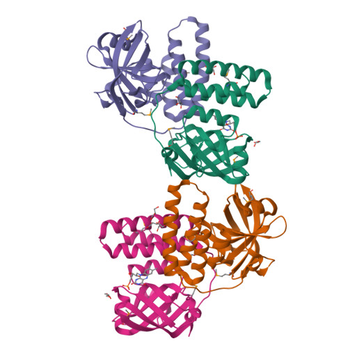
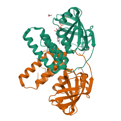
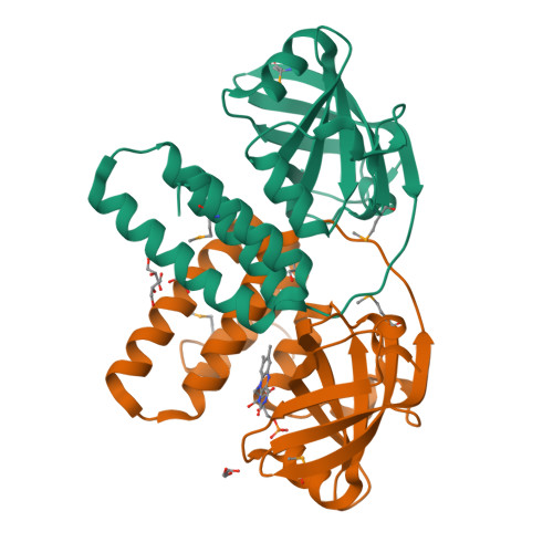
Crystal structure of a hypothetical protein from Archaeoglobus fulgidus binding riboflavin 5'-phosphate
Bonanno, J.B., Dickey, M., Bain, K.T., Adams, J., Ozyurt, S., Wasserman, S., Sauder, J.M., Burley, S.K., Almo, S.C.To be published.
Experimental Data Snapshot
Entity ID: 1 | |||||
|---|---|---|---|---|---|
| Molecule | Chains | Sequence Length | Organism | Details | Image |
| Hypothetical protein | 199 | Archaeoglobus fulgidus | Mutation(s): 4 Gene Names: AF_1834 | 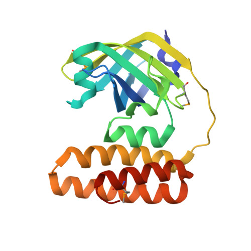 | |
UniProt | |||||
Find proteins for O28442 (Archaeoglobus fulgidus (strain ATCC 49558 / DSM 4304 / JCM 9628 / NBRC 100126 / VC-16)) Explore O28442 Go to UniProtKB: O28442 | |||||
Entity Groups | |||||
| Sequence Clusters | 30% Identity50% Identity70% Identity90% Identity95% Identity100% Identity | ||||
| UniProt Group | O28442 | ||||
Sequence AnnotationsExpand | |||||
| |||||
| Ligands 2 Unique | |||||
|---|---|---|---|---|---|
| ID | Chains | Name / Formula / InChI Key | 2D Diagram | 3D Interactions | |
| FMN Query on FMN | E [auth A], M [auth D] | FLAVIN MONONUCLEOTIDE C17 H21 N4 O9 P FVTCRASFADXXNN-SCRDCRAPSA-N |  | ||
| GOL Query on GOL | F [auth A] G [auth A] H [auth A] I [auth B] J [auth B] | GLYCEROL C3 H8 O3 PEDCQBHIVMGVHV-UHFFFAOYSA-N |  | ||
| Modified Residues 1 Unique | |||||
|---|---|---|---|---|---|
| ID | Chains | Type | Formula | 2D Diagram | Parent |
| MSE Query on MSE | A, B, C, D | L-PEPTIDE LINKING | C5 H11 N O2 Se |  | MET |
| Length ( Å ) | Angle ( ˚ ) |
|---|---|
| a = 48.042 | α = 90 |
| b = 103.182 | β = 97.01 |
| c = 103.842 | γ = 90 |
| Software Name | Purpose |
|---|---|
| REFMAC | refinement |
| CBASS | data collection |
| DENZO | data reduction |
| SCALEPACK | data scaling |
| SHELXD | phasing |
RCSB PDB is hosted by

RCSB PDB is a member of the