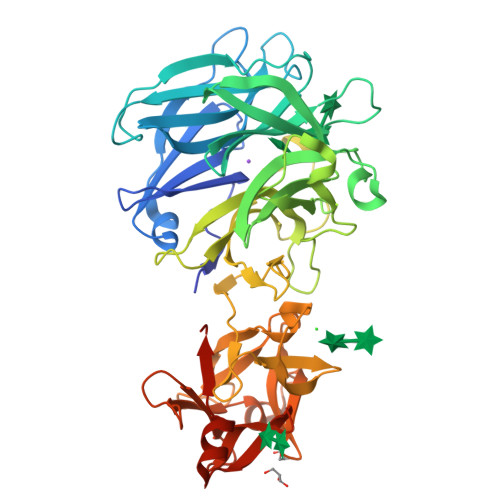
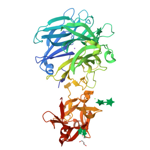
3AKH | pdb_00003akh

Crystal structure of exo-1,5-alpha-L-arabinofuranosidase complexed with alpha-1,5-L-arabinofuranotriose


Experimental Data Snapshot

  • Method: X-RAY DIFFRACTION
  • Resolution: 1.70 Å
  • R-Value Free: 
    0.220 (Depositor), 0.220 (DCC) 
  • R-Value Work: 
    0.188 (Depositor), 0.190 (DCC) 
  • R-Value Observed: 
    0.190 (Depositor) 

Starting Model: experimental
View more details

wwPDB Validation   3D Report Full Report


Ligand Structure Quality Assessment 

Created with Raphaël 2.3.0Worse 01 BetterLigand structure goodness of fit to experimental dataBest fitted AHRClick on this verticalbar to view details

This is version 2.1 of the entry. See complete history


Literature

Crystal Structure of an Exo-1,5-{alpha}-L-arabinofuranosidase from Streptomyces avermitilis Provides Insights into the Mechanism of Substrate Discrimination between Exo- and Endo-type Enzymes in Glycoside Hydrolase Family 43.

Fujimoto, Z.Ichinose, H.Maehara, T.Honda, M.Kitaoka, M.Kaneko, S.

(2010) J Biological Chem 285: 34134-34143

  • DOI: https://doi.org/10.1074/jbc.M110.164251
  • Primary Citation of Related Structures:  
    3AKF, 3AKG, 3AKH, 3AKI

  • PubMed Abstract: 

    Exo-1,5-α-L-arabinofuranosidases belonging to glycoside hydrolase family 43 have strict substrate specificity. These enzymes hydrolyze only the α-1,5-linkages of linear arabinan and arabino-oligosaccharides in an exo-acting manner. The enzyme from Streptomyces avermitilis contains a core catalytic domain belonging to glycoside hydrolase family 43 and a C-terminal arabinan binding module belonging to carbohydrate binding module family 42. We determined the crystal structure of intact exo-1,5-α-L-arabinofuranosidase. The catalytic module is composed of a 5-bladed β-propeller topologically identical to the other family 43 enzymes. The arabinan binding module had three similar subdomains assembled against one another around a pseudo-3-fold axis, forming a β-trefoil-fold. A sugar complex structure with α-1,5-L-arabinofuranotriose revealed three subsites in the catalytic domain, and a sugar complex structure with α-L-arabinofuranosyl azide revealed three arabinose-binding sites in the carbohydrate binding module. A mutagenesis study revealed that substrate specificity was regulated by residues Asn-159, Tyr-192, and Leu-289 located at the aglycon side of the substrate-binding pocket. The exo-acting manner of the enzyme was attributed to the strict pocket structure of subsite -1, formed by the flexible loop region Tyr-281-Arg-294 and the side chain of Tyr-40, which occupied the positions corresponding to the catalytic glycon cleft of GH43 endo-acting enzymes.


  • Organizational Affiliation

    Protein Research Unit, National Institute of Agrobiological Sciences, 2-1-2 Kannondai, Tsukuba, Ibaraki 305-8602, Japan.


Macromolecules
Find similar proteins by:  (by identity cutoff)  |  3D Structure
Entity ID: 1
MoleculeChains Sequence LengthOrganismDetailsImage
Putative secreted alpha L-arabinofuranosidase II468Streptomyces avermitilis MA-4680 = NBRC 14893Mutation(s): 0 
Gene Names: abfASAV1043SAV_1043
EC: 3.2.1.55
UniProt
Find proteins for Q82P90 (Streptomyces avermitilis (strain ATCC 31267 / DSM 46492 / JCM 5070 / NBRC 14893 / NCIMB 12804 / NRRL 8165 / MA-4680))
Explore Q82P90 
Go to UniProtKB:  Q82P90
Entity Groups  
Sequence Clusters30% Identity50% Identity70% Identity90% Identity95% Identity100% Identity
UniProt GroupQ82P90
Sequence Annotations
Expand
  • Reference Sequence
Oligosaccharides

Help

Entity ID: 2
MoleculeChains Length2D Diagram Glycosylation3D Interactions
alpha-L-arabinofuranose-(1-5)-alpha-L-arabinofuranose
B, C, D
2N/A
Glycosylation Resources
GlyTouCan:  G41001SE
GlyCosmos:  G41001SE
Experimental Data & Validation

Experimental Data

  • Method: X-RAY DIFFRACTION
  • Resolution: 1.70 Å
  • R-Value Free:  0.220 (Depositor), 0.220 (DCC) 
  • R-Value Work:  0.188 (Depositor), 0.190 (DCC) 
  • R-Value Observed: 0.190 (Depositor) 
Space Group: P 21 21 21
Unit Cell:
Length ( Å )Angle ( ˚ )
a = 41.237α = 90
b = 91.341β = 90
c = 135.962γ = 90
Software Package:
Software NamePurpose
SERGUIdata collection
MOLREPphasing
REFMACrefinement
HKL-2000data reduction
SCALEPACKdata scaling

Structure Validation

View Full Validation Report


Ligand Structure Quality Assessment 

Created with Raphaël 2.3.0Worse 01 BetterLigand structure goodness of fit to experimental dataBest fitted AHRClick on this verticalbar to view details

Entry History 

Deposition Data

Revision History  (Full details and data files)

  • Version 1.0: 2010-08-25
    Type: Initial release
  • Version 1.1: 2011-07-13
    Changes: Version format compliance
  • Version 1.2: 2017-10-11
    Changes: Refinement description
  • Version 2.0: 2020-07-29
    Type: Remediation
    Reason: Carbohydrate remediation
    Changes: Atomic model, Data collection, Database references, Derived calculations, Structure summary
  • Version 2.1: 2023-11-01
    Changes: Data collection, Database references, Derived calculations, Refinement description, Structure summary