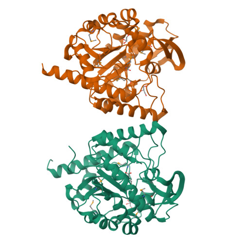
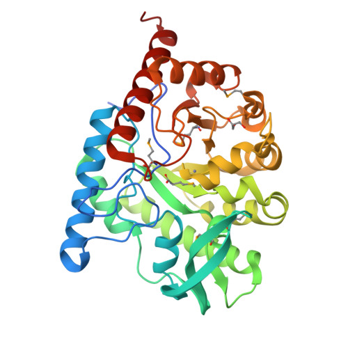
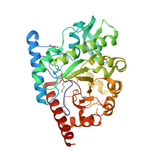
The structure of a B12-independent methionine synthase from Shewanella sp. W3-18-1 in complex with Selenomethionine.
Cuff, M.E., Li, H., Hatzos-Skintges, C., Tesar, C., Bearden, J., Clancy, A., Joachimiak, A.To be published.
Experimental Data Snapshot
Entity ID: 1 | |||||
|---|---|---|---|---|---|
| Molecule | Chains | Sequence Length | Organism | Details | Image |
| Methionine synthase (B12-independent) | 357 | Shewanella sp. W3-18-1 | Mutation(s): 0 Gene Names: Sputw3181_1250 EC: 2.1.1.14 | 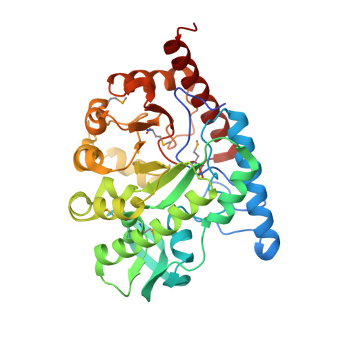 | |
Entity Groups | |||||
| Sequence Clusters | 30% Identity50% Identity70% Identity90% Identity95% Identity100% Identity | ||||
Sequence AnnotationsExpand | |||||
| |||||
| Ligands 3 Unique | |||||
|---|---|---|---|---|---|
| ID | Chains | Name / Formula / InChI Key | 2D Diagram | 3D Interactions | |
| MSE Query on MSE | D [auth A], G [auth B] | SELENOMETHIONINE C5 H11 N O2 Se RJFAYQIBOAGBLC-BYPYZUCNSA-N |  | ||
| GOL Query on GOL | E [auth A], H [auth B], I [auth B] | GLYCEROL C3 H8 O3 PEDCQBHIVMGVHV-UHFFFAOYSA-N |  | ||
| ZN Query on ZN | C [auth A], F [auth B] | ZINC ION Zn PTFCDOFLOPIGGS-UHFFFAOYSA-N |  | ||
| Modified Residues 1 Unique | |||||
|---|---|---|---|---|---|
| ID | Chains | Type | Formula | 2D Diagram | Parent |
| MSE Query on MSE | A, B | L-PEPTIDE LINKING | C5 H11 N O2 Se | | MET |
| Length ( Å ) | Angle ( ˚ ) |
|---|---|
| a = 46.099 | α = 90 |
| b = 89.371 | β = 92.45 |
| c = 86.526 | γ = 90 |
| Software Name | Purpose |
|---|---|
| DENZO | data reduction |
| SCALEPACK | data scaling |
| MLPHARE | phasing |
| DM | phasing |
| REFMAC | refinement |
| PDB_EXTRACT | data extraction |
| SBC-Collect | data collection |
| HKL-3000 | data reduction |
| HKL-3000 | data scaling |
| HKL-3000 | phasing |
| SHELXD | phasing |
| SHELXE | model building |
| SOLVE | phasing |
| RESOLVE | phasing |
| ARP/wARP | model building |
| CCP4 | phasing |
| O | model building |
| Coot | model building |
RCSB PDB is hosted by

RCSB PDB is a member of the