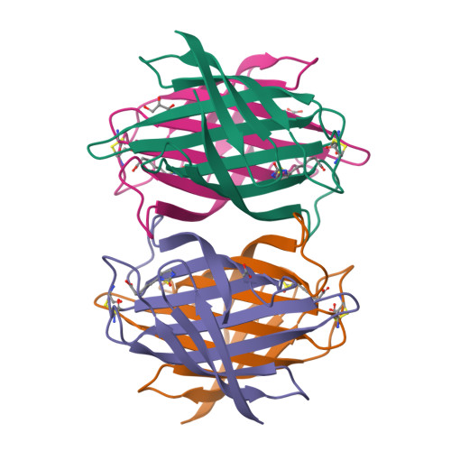
Crystal Structure of Core-Bradavidin
Airenne, T.T., Maatta, J.A.E., Nordlund, H., Kulomaa, M.S., Johnson, M.S.To be published.
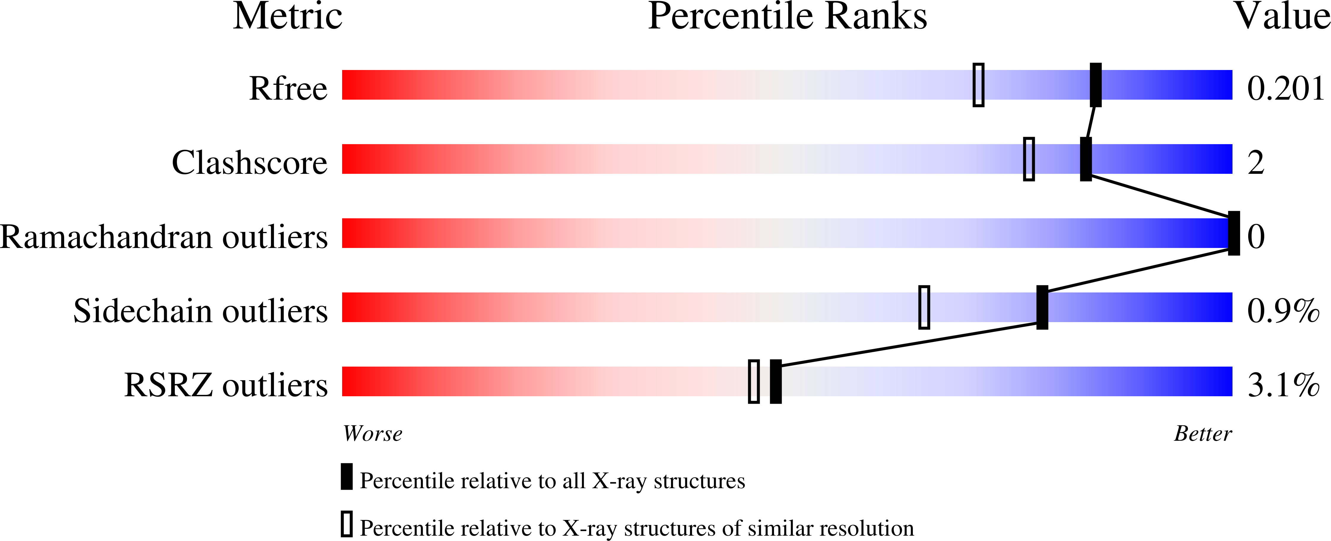
Experimental Data Snapshot
Starting Model: experimental
View more details
Entity ID: 1 | |||||
|---|---|---|---|---|---|
| Molecule | Chains | Sequence Length | Organism | Details | Image |
| BLR5658 PROTEIN | 118 | Bradyrhizobium japonicum | Mutation(s): 0 | 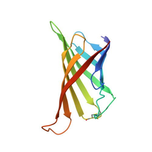 | |
UniProt | |||||
Find proteins for Q89IH6 (Bradyrhizobium diazoefficiens (strain JCM 10833 / BCRC 13528 / IAM 13628 / NBRC 14792 / USDA 110)) Explore Q89IH6 Go to UniProtKB: Q89IH6 | |||||
Entity Groups | |||||
| Sequence Clusters | 30% Identity50% Identity70% Identity90% Identity95% Identity100% Identity | ||||
| UniProt Group | Q89IH6 | ||||
Sequence AnnotationsExpand | |||||
| |||||
| Ligands 3 Unique | |||||
|---|---|---|---|---|---|
| ID | Chains | Name / Formula / InChI Key | 2D Diagram | 3D Interactions | |
| BTN Query on BTN | E [auth A], G [auth B], I [auth C], K [auth D] | BIOTIN C10 H16 N2 O3 S YBJHBAHKTGYVGT-ZKWXMUAHSA-N |  | ||
| GOL Query on GOL | F [auth A] | GLYCEROL C3 H8 O3 PEDCQBHIVMGVHV-UHFFFAOYSA-N |  | ||
| ACT Query on ACT | H [auth B], J [auth C], L [auth D] | ACETATE ION C2 H3 O2 QTBSBXVTEAMEQO-UHFFFAOYSA-M |  | ||
| Length ( Å ) | Angle ( ˚ ) |
|---|---|
| a = 49.946 | α = 90 |
| b = 78.647 | β = 90 |
| c = 100.12 | γ = 90 |
| Software Name | Purpose |
|---|---|
| REFMAC | refinement |
| XDS | data reduction |
| PHASER | phasing |
RCSB PDB is hosted by

RCSB PDB is a member of the