4HEU | pdb_00004heu

Crystal Structure of PDE10A with a biaryl ether inhibitor ((1-(3-(4-((1H-benzo[d]imidazol-2-yl)amino)phenoxy)pyridin-2-yl)piperidin-4-yl)methanol)


Experimental Data Snapshot

  • Method: X-RAY DIFFRACTION
  • Resolution: 2.00 Å
  • R-Value Free: 
    0.199 (Depositor), 0.197 (DCC) 
  • R-Value Work: 
    0.186 (Depositor), 0.184 (DCC) 
  • R-Value Observed: 
    0.187 (Depositor) 

wwPDB Validation   3D Report Full Report


Ligand Structure Quality Assessment 

Created with Raphaël 2.3.0Worse 01 BetterLigand structure goodness of fit to experimental dataBest fitted 15JClick on this verticalbar to view details

This is version 1.1 of the entry. See complete history


Literature

Discovery of selective biaryl ethers as PDE10A inhibitors: Improvement in potency and mitigation of Pgp-mediated efflux.

Rzasa, R.M.Hu, E.Rumfelt, S.Chen, N.Andrews, K.L.Chmait, S.Falsey, J.R.Zhong, W.Jones, A.D.Porter, A.Louie, S.W.Zhao, X.Treanor, J.J.Allen, J.R.

(2012) Bioorg Med Chem Lett 22: 7371-7375

  • DOI: https://doi.org/10.1016/j.bmcl.2012.10.078
  • Primary Citation of Related Structures:  
    4HEU, 4HF4

  • PubMed Abstract: 

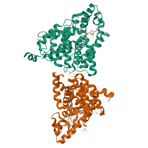
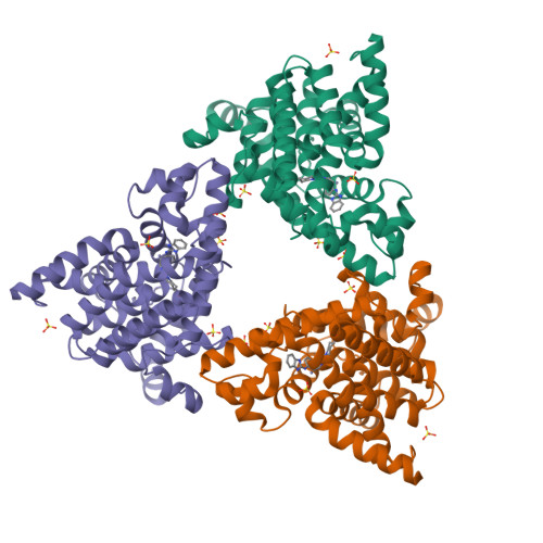
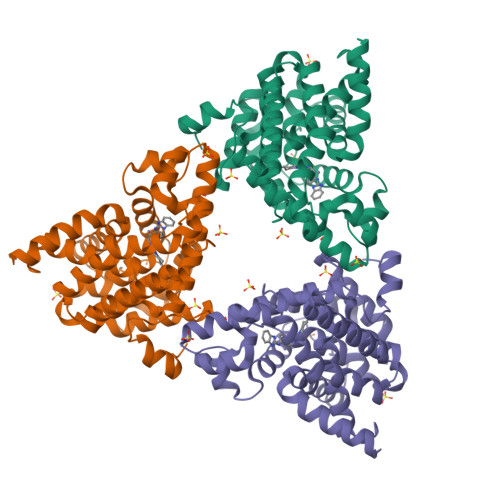
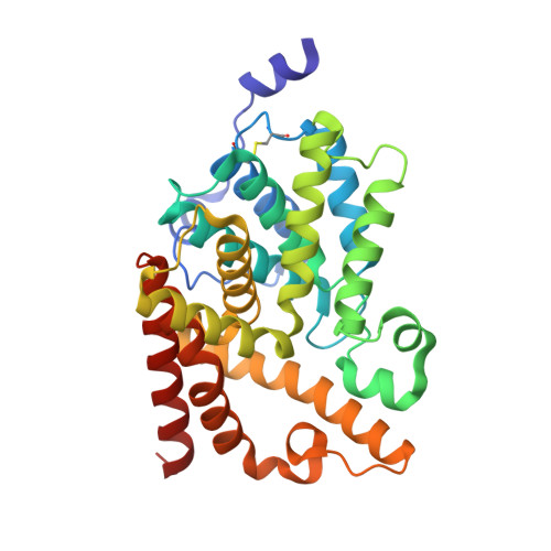
    We report the discovery of a novel series of biaryl ethers as potent and selective PDE10A inhibitors. Structure-activity studies improved the potency and decreased Pgp-mediated efflux found in the initial compound 4. X-ray crystallographic studies revealed two novel binding modes to the catalytic site of the PDE10A enzyme.


  • Organizational Affiliation

    Department of Medicinal Chemistry, Amgen Inc., One Amgen Center Drive, Thousand Oaks, CA 91320-1799, USA. rrzasa@amgen.com


Macromolecules
Find similar proteins by:  (by identity cutoff)  |  3D Structure
Entity ID: 1
MoleculeChains Sequence LengthOrganismDetailsImage
cAMP and cAMP-inhibited cGMP 3',5'-cyclic phosphodiesterase 10A
A, B
318Homo sapiensMutation(s): 0 
Gene Names: PDE10A
EC: 3.1.4.35 (PDB Primary Data), 3.1.4.17 (UniProt)
UniProt & NIH Common Fund Data Resources
Find proteins for Q9Y233 (Homo sapiens)
Explore Q9Y233 
Go to UniProtKB:  Q9Y233
PHAROS:  Q9Y233
GTEx:  ENSG00000112541 
Entity Groups  
Sequence Clusters30% Identity50% Identity70% Identity90% Identity95% Identity100% Identity
UniProt GroupQ9Y233
Sequence Annotations
Expand
  • Reference Sequence
Small Molecules
Ligands 3 Unique
IDChains Name / Formula / InChI Key2D Diagram3D Interactions
15J
Query on 15J

Download Ideal Coordinates CCD File 
K [auth A],
T [auth B]
(1-{3-[4-(1H-benzimidazol-2-ylamino)phenoxy]pyridin-2-yl}piperidin-4-yl)methanol
C24 H25 N5 O2
XGFBQNCOZRNUGL-UHFFFAOYSA-N
SO4
Query on SO4

Download Ideal Coordinates CCD File 
E [auth A]
F [auth A]
G [auth A]
H [auth A]
I [auth A]
SULFATE ION
O4 S
QAOWNCQODCNURD-UHFFFAOYSA-L
ZN
Query on ZN

Download Ideal Coordinates CCD File 
C [auth A],
D [auth A],
L [auth B],
M [auth B]
ZINC ION
Zn
PTFCDOFLOPIGGS-UHFFFAOYSA-N
Experimental Data & Validation

Experimental Data

  • Method: X-RAY DIFFRACTION
  • Resolution: 2.00 Å
  • R-Value Free:  0.199 (Depositor), 0.197 (DCC) 
  • R-Value Work:  0.186 (Depositor), 0.184 (DCC) 
  • R-Value Observed: 0.187 (Depositor) 
Space Group: F 2 3
Unit Cell:
Length ( Å )Angle ( ˚ )
a = 252.859α = 90
b = 252.859β = 90
c = 252.859γ = 90
Software Package:
Software NamePurpose
ADSCdata collection
MOLREPphasing
REFMACrefinement
HKL-2000data reduction
SCALEPACKdata scaling

Structure Validation

View Full Validation Report


Ligand Structure Quality Assessment 

Created with Raphaël 2.3.0Worse 01 BetterLigand structure goodness of fit to experimental dataBest fitted 15JClick on this verticalbar to view details

Entry History 

Deposition Data

Revision History  (Full details and data files)

  • Version 1.0: 2012-12-12
    Type: Initial release
  • Version 1.1: 2024-11-27
    Changes: Data collection, Database references, Derived calculations, Structure summary