6PXI | pdb_00006pxi

The crystal structure of a singly capped HslUV complex with an axial pore plug and a HslU E257Q mutation


Experimental Data Snapshot

  • Method: X-RAY DIFFRACTION
  • Resolution: 3.45 Å
  • R-Value Free: 
    0.285 (Depositor), 0.260 (DCC) 
  • R-Value Work: 
    0.257 (Depositor), 0.240 (DCC) 
  • R-Value Observed: 
    0.261 (Depositor) 

Starting Model: experimental
View more details

wwPDB Validation   3D Report Full Report


Ligand Structure Quality Assessment 

Created with Raphaël 2.3.0Worse 01 BetterLigand structure goodness of fit to experimental dataBest fitted ADPClick on this verticalbar to view details

This is version 1.3 of the entry. See complete history


Literature

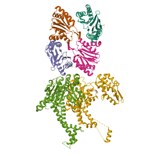
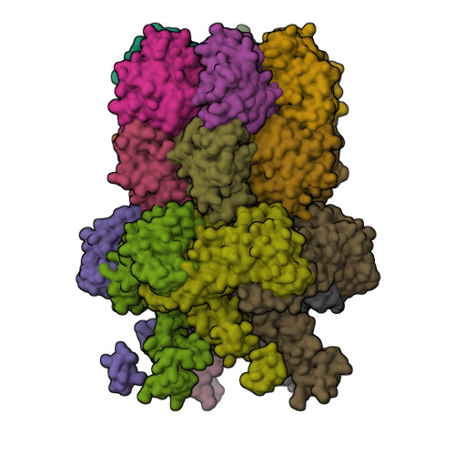
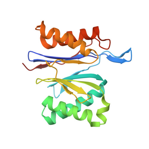
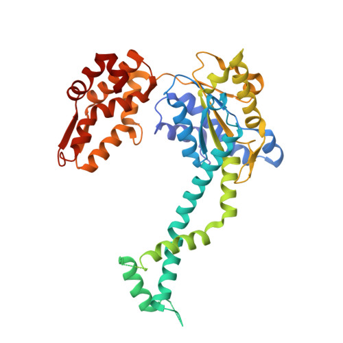
Heat activates the AAA+ HslUV protease by melting an axial autoinhibitory plug.

Baytshtok, V.Fei, X.Shih, T.T.Grant, R.A.Santos, J.C.Baker, T.A.Sauer, R.T.

(2021) Cell Rep 34: 108639-108639

  • DOI: https://doi.org/10.1016/j.celrep.2020.108639
  • Primary Citation of Related Structures:  
    6PXI, 6PXK, 6PXL

  • PubMed Abstract: 

    At low temperatures, protein degradation by the AAA+ HslUV protease is very slow. New crystal structures reveal that residues in the intermediate domain of the HslU 6 unfoldase can plug its axial channel, blocking productive substrate binding and subsequent unfolding, translocation, and degradation by the HslV 12 peptidase. Biochemical experiments with wild-type and mutant enzymes support a model in which heat-induced melting of this autoinhibitory plug activates HslUV proteolysis.


  • Organizational Affiliation

    Department of Biology, Massachusetts Institute of Technology, Cambridge, MA 02139, USA.


Macromolecules
Find similar proteins by:  (by identity cutoff)  |  3D Structure
Entity ID: 1
MoleculeChains Sequence LengthOrganismDetailsImage
ATP-dependent protease subunit HslV
A, B, C, D
174Escherichia coliMutation(s): 0 
Gene Names: hslVhtpOyiiCb3932JW3903
EC: 3.4.25.2
UniProt
Find proteins for P0A7B8 (Escherichia coli (strain K12))
Explore P0A7B8 
Go to UniProtKB:  P0A7B8
Entity Groups  
Sequence Clusters30% Identity50% Identity70% Identity90% Identity95% Identity100% Identity
UniProt GroupP0A7B8
Sequence Annotations
Expand
  • Reference Sequence
Find similar proteins by:  (by identity cutoff)  |  3D Structure
Entity ID: 2
MoleculeChains Sequence LengthOrganismDetailsImage
ATP-dependent protease ATPase subunit HslU
E, F
448Escherichia coliMutation(s): 1 
Gene Names: hslUhtpIb3931JW3902
UniProt
Find proteins for P0A6H5 (Escherichia coli (strain K12))
Explore P0A6H5 
Go to UniProtKB:  P0A6H5
Entity Groups  
Sequence Clusters30% Identity50% Identity70% Identity90% Identity95% Identity100% Identity
UniProt GroupP0A6H5
Sequence Annotations
Expand
  • Reference Sequence
Experimental Data & Validation

Experimental Data

  • Method: X-RAY DIFFRACTION
  • Resolution: 3.45 Å
  • R-Value Free:  0.285 (Depositor), 0.260 (DCC) 
  • R-Value Work:  0.257 (Depositor), 0.240 (DCC) 
  • R-Value Observed: 0.261 (Depositor) 
Space Group: P 3 2 1
Unit Cell:
Length ( Å )Angle ( ˚ )
a = 168.604α = 90
b = 168.604β = 90
c = 162.886γ = 120
Software Package:
Software NamePurpose
HKL-2000data scaling
PHENIXrefinement
PDB_EXTRACTdata extraction
HKL-2000data reduction
PHASERphasing

Structure Validation

View Full Validation Report


Ligand Structure Quality Assessment 

Created with Raphaël 2.3.0Worse 01 BetterLigand structure goodness of fit to experimental dataBest fitted ADPClick on this verticalbar to view details

Entry History & Funding Information

Deposition Data


Funding OrganizationLocationGrant Number
National Institutes of Health/National Human Genome Research Institute (NIH/NHGRI)United States--

Revision History  (Full details and data files)

  • Version 1.0: 2020-07-29
    Type: Initial release
  • Version 1.1: 2021-02-10
    Changes: Database references
  • Version 1.2: 2023-10-11
    Changes: Data collection, Database references, Refinement description
  • Version 1.3: 2024-06-19
    Changes: Database references, Source and taxonomy