7YRK | pdb_00007yrk

Crystal structure of the hen egg lysozyme-2-oxidobenzylidene-threoninato-copper (II) complex


Experimental Data Snapshot

  • Method: X-RAY DIFFRACTION
  • Resolution: 0.92 Å
  • R-Value Free: 
    0.128 (Depositor), 0.126 (DCC) 
  • R-Value Work: 
    0.113 (Depositor), 0.111 (DCC) 
  • R-Value Observed: 
    0.114 (Depositor) 

Starting Model: experimental
View more details

wwPDB Validation   3D Report Full Report


Ligand Structure Quality Assessment 

Created with Raphaël 2.3.0Worse 01 BetterLigand structure goodness of fit to experimental dataBest fitted JI6Click on this verticalbar to view details

This is version 2.0 of the entry. See complete history


Literature

A novel hybrid protein composed of superoxide-dismutase-active Cu(II) complex and lysozyme.

Furuya, T.Nakane, D.Kitanishi, K.Katsuumi, N.Tsaturyan, A.Shcherbakov, I.N.Unno, M.Akitsu, T.

(2023) Sci Rep 13: 6892-6892

  • DOI: https://doi.org/10.1038/s41598-023-33926-1
  • Primary Citation of Related Structures:  
    7YRK

  • PubMed Abstract: 

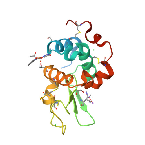
    A novel hybrid protein composed of a superoxide dismutase-active Cu(II) complex (CuST) and lysozyme (CuST@lysozyme) was prepared. The results of the spectroscopic and electrochemical analyses confirmed that CuST binds to lysozyme. We determined the crystal structure of CuST@lysozyme at 0.92 Å resolution, which revealed that the His15 imidazole group of lysozyme binds to the Cu(II) center of CuST in the equatorial position. In addition, CuST was fixed in position by the weak axial coordination of the Thr89 hydroxyl group and the hydrogen bond between the guanidinium group of the Arg14 residue and the hydroxyl group of CuST. Furthermore, the combination of CuST with lysozyme did not decrease the superoxide dismutase activity of CuST. Based on the spectral, electrochemical, structural studies, and quantum chemical calculations, an O 2 - disproportionation mechanism catalyzed by CuST@lysozyme is proposed.


  • Organizational Affiliation

    Department of Chemistry, Faculty of Science, Tokyo University of Science, 1-3 Kagurazaka, Shinjuku-ku, Tokyo, 162-8601, Japan.


Macromolecules
Find similar proteins by:  (by identity cutoff)  |  3D Structure
Entity ID: 1
MoleculeChains Sequence LengthOrganismDetailsImage
Lysozyme C147Gallus gallusMutation(s): 0 
EC: 3.2.1.17
UniProt
Find proteins for P00698 (Gallus gallus)
Explore P00698 
Go to UniProtKB:  P00698
Entity Groups  
Sequence Clusters30% Identity50% Identity70% Identity90% Identity95% Identity100% Identity
UniProt GroupP00698
Sequence Annotations
Expand
  • Reference Sequence
Small Molecules
Ligands 4 Unique
IDChains Name / Formula / InChI Key2D Diagram3D Interactions
JI6 (Subject of Investigation/LOI)
Query on JI6

Download Ideal Coordinates CCD File 
D [auth A](6~{S})-6-[(1~{R})-1-oxidanylethyl]-2,4-dioxa-7$l^{4}-aza-3$l^{3}-cupratricyclo[7.4.0.0^{3,7}]trideca-1(13),7,9,11-tetraen-5-one
C11 H11 Cu N O4
RLMUPHKMYNMLMB-NBDMYRNNSA-L
EDO
Query on EDO

Download Ideal Coordinates CCD File 
B [auth A],
H [auth A]
1,2-ETHANEDIOL
C2 H6 O2
LYCAIKOWRPUZTN-UHFFFAOYSA-N
CL
Query on CL

Download Ideal Coordinates CCD File 
C [auth A],
E [auth A],
F [auth A],
G [auth A]
CHLORIDE ION
Cl
VEXZGXHMUGYJMC-UHFFFAOYSA-M
NA
Query on NA

Download Ideal Coordinates CCD File 
I [auth A],
J [auth A],
K [auth A]
SODIUM ION
Na
FKNQFGJONOIPTF-UHFFFAOYSA-N
Modified Residues  1 Unique
IDChains TypeFormula2D DiagramParent
MEN
Query on MEN
A
L-PEPTIDE LINKINGC5 H10 N2 O3ASN
Experimental Data & Validation

Experimental Data

  • Method: X-RAY DIFFRACTION
  • Resolution: 0.92 Å
  • R-Value Free:  0.128 (Depositor), 0.126 (DCC) 
  • R-Value Work:  0.113 (Depositor), 0.111 (DCC) 
  • R-Value Observed: 0.114 (Depositor) 
Space Group: P 43 21 2
Unit Cell:
Length ( Å )Angle ( ˚ )
a = 78.671α = 90
b = 78.671β = 90
c = 37.203γ = 90
Software Package:
Software NamePurpose
PHENIXrefinement
XDSdata reduction
XDSdata scaling
REFMACphasing

Structure Validation

View Full Validation Report


Ligand Structure Quality Assessment 

Created with Raphaël 2.3.0Worse 01 BetterLigand structure goodness of fit to experimental dataBest fitted JI6Click on this verticalbar to view details

Entry History & Funding Information

Deposition Data


Funding OrganizationLocationGrant Number
Japan Society for the Promotion of Science (JSPS)Japan20H00336

Revision History  (Full details and data files)

  • Version 1.0: 2023-05-10
    Type: Initial release
  • Version 1.1: 2023-11-29
    Changes: Data collection, Refinement description
  • Version 2.0: 2024-09-25
    Changes: Atomic model, Author supporting evidence, Data collection, Derived calculations, Non-polymer description, Polymer sequence, Structure summary