7ZZU | pdb_00007zzu

Inhibitory Ligand binding to HDAC2


Experimental Data Snapshot

  • Method: X-RAY DIFFRACTION
  • Resolution: 1.85 Å
  • R-Value Free: 
    0.182 (Depositor), 0.190 (DCC) 
  • R-Value Work: 
    0.154 (Depositor), 0.170 (DCC) 
  • R-Value Observed: 
    0.155 (Depositor) 

wwPDB Validation   3D Report Full Report


Ligand Structure Quality Assessment 

Created with Raphaël 2.3.0Worse 01 BetterLigand structure goodness of fit to experimental dataBest fitted KKIClick on this verticalbar to view detailsBest fitted NHEClick on this verticalbar to view details

This is version 1.2 of the entry. See complete history


Literature

Fragment-Based Discovery of a Novel, Brain Penetrant, Orally Active HDAC2 Inhibitor.

Tamanini, E.Miyamura, S.Buck, I.M.Cons, B.D.Dawson, L.East, C.Futamura, T.Goto, S.Griffiths-Jones, C.Hashimoto, T.Heightman, T.D.Ishikawa, S.Ito, H.Kaneko, Y.Kawato, T.Kondo, K.Kurihara, N.McCarthy, J.M.Mori, Y.Nagase, T.Nakaishi, Y.Reeks, J.Sato, A.Schopf, P.Tai, K.Tamai, T.Tisi, D.Woolford, A.J.

(2022) ACS Med Chem Lett 13: 1591-1597

  • DOI: https://doi.org/10.1021/acsmedchemlett.2c00272
  • Primary Citation of Related Structures:  
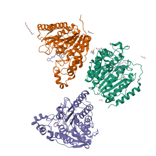
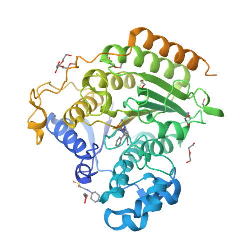
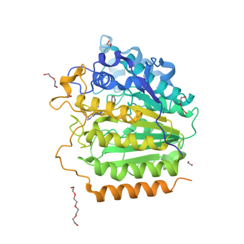
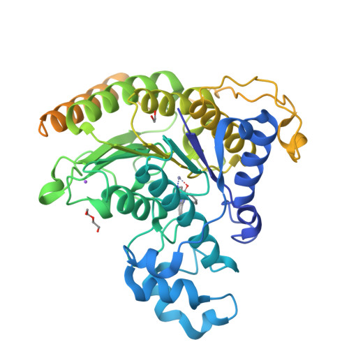
    7ZZO, 7ZZP, 7ZZR, 7ZZS, 7ZZT, 7ZZU, 7ZZW, 8A0B

  • PubMed Abstract: 

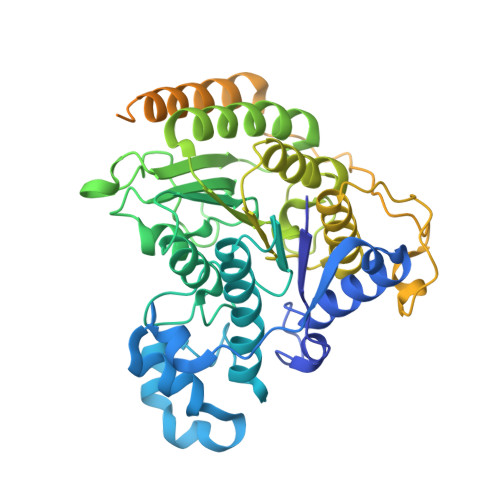
    Fragment-based ligand discovery was successfully applied to histone deacetylase HDAC2. In addition to the anticipated hydroxamic acid- and benzamide-based fragment screening hits, a low affinity (∼1 mM) α-amino-amide zinc binding fragment was identified, as well as fragments binding to other regions of the catalytic site. This alternative zinc-binding fragment was further optimized, guided by the structural information from protein-ligand complex X-ray structures, into a sub-μM, brain penetrant, HDAC2 inhibitor ( 17 ) capable of modulating histone acetylation levels in vivo .


  • Organizational Affiliation

    Astex Pharmaceuticals, 436 Cambridge Science Park, Milton Road, Cambridge CB4 0QA, U.K.


Macromolecules
Find similar proteins by:  (by identity cutoff)  |  3D Structure
Entity ID: 1
MoleculeChains Sequence LengthOrganismDetailsImage
Histone deacetylase 2
A, B, C
498Homo sapiensMutation(s): 0 
Gene Names: HDAC2
EC: 3.5.1.98 (PDB Primary Data), 3.5.1 (PDB Primary Data)
UniProt & NIH Common Fund Data Resources
Find proteins for Q92769 (Homo sapiens)
Explore Q92769 
Go to UniProtKB:  Q92769
PHAROS:  Q92769
GTEx:  ENSG00000196591 
Entity Groups  
Sequence Clusters30% Identity50% Identity70% Identity90% Identity95% Identity100% Identity
UniProt GroupQ92769
Sequence Annotations
Expand
  • Reference Sequence
Small Molecules
Ligands 8 Unique
IDChains Name / Formula / InChI Key2D Diagram3D Interactions
KKI (Subject of Investigation/LOI)
Query on KKI

Download Ideal Coordinates CCD File 
AA [auth B],
GA [auth C],
P [auth A]
2-[4-[(2~{R},4~{S})-4-phenylpyrrolidin-2-yl]carbonylpiperazin-1-yl]pyridine-3-carbonitrile
C21 H23 N5 O
ZOCRTPMGIHWFKT-RTBURBONSA-N
NHE (Subject of Investigation/LOI)
Query on NHE

Download Ideal Coordinates CCD File 
Q [auth A]2-[N-CYCLOHEXYLAMINO]ETHANE SULFONIC ACID
C8 H17 N O3 S
MKWKNSIESPFAQN-UHFFFAOYSA-N
PG4
Query on PG4

Download Ideal Coordinates CCD File 
F [auth A],
T [auth B]
TETRAETHYLENE GLYCOL
C8 H18 O5
UWHCKJMYHZGTIT-UHFFFAOYSA-N
PEG
Query on PEG

Download Ideal Coordinates CCD File 
BA [auth C]
G [auth A]
I [auth A]
J [auth A]
S [auth B]
DI(HYDROXYETHYL)ETHER
C4 H10 O3
MTHSVFCYNBDYFN-UHFFFAOYSA-N
ZN
Query on ZN

Download Ideal Coordinates CCD File 
DA [auth C],
M [auth A],
X [auth B]
ZINC ION
Zn
PTFCDOFLOPIGGS-UHFFFAOYSA-N
EDO
Query on EDO

Download Ideal Coordinates CCD File 
CA [auth C]
D [auth A]
E [auth A]
H [auth A]
K [auth A]
1,2-ETHANEDIOL
C2 H6 O2
LYCAIKOWRPUZTN-UHFFFAOYSA-N
CA
Query on CA

Download Ideal Coordinates CCD File 
EA [auth C],
N [auth A],
Y [auth B]
CALCIUM ION
Ca
BHPQYMZQTOCNFJ-UHFFFAOYSA-N
NA
Query on NA

Download Ideal Coordinates CCD File 
FA [auth C],
O [auth A],
Z [auth B]
SODIUM ION
Na
FKNQFGJONOIPTF-UHFFFAOYSA-N
Binding Affinity Annotations 
IDSourceBinding Affinity
KKI BindingDB:  7ZZU IC50: 1800 (nM) from 1 assay(s)
Experimental Data & Validation

Experimental Data

  • Method: X-RAY DIFFRACTION
  • Resolution: 1.85 Å
  • R-Value Free:  0.182 (Depositor), 0.190 (DCC) 
  • R-Value Work:  0.154 (Depositor), 0.170 (DCC) 
  • R-Value Observed: 0.155 (Depositor) 
Space Group: P 21 21 21
Unit Cell:
Length ( Å )Angle ( ˚ )
a = 92.35α = 90
b = 98.352β = 90
c = 139.291γ = 90
Software Package:
Software NamePurpose
REFMACrefinement
PDB_EXTRACTdata extraction
HKL-2000data reduction
Aimlessdata scaling
REFMACphasing

Structure Validation

View Full Validation Report


Ligand Structure Quality Assessment 

Created with Raphaël 2.3.0Worse 01 BetterLigand structure goodness of fit to experimental dataBest fitted KKIClick on this verticalbar to view detailsBest fitted NHEClick on this verticalbar to view details

Entry History & Funding Information

Deposition Data


Funding OrganizationLocationGrant Number
Other privateUnited Kingdom--

Revision History  (Full details and data files)

  • Version 1.0: 2022-09-21
    Type: Initial release
  • Version 1.1: 2022-11-02
    Changes: Database references
  • Version 1.2: 2024-06-19
    Changes: Data collection