1AZT

GS-ALPHA COMPLEXED WITH GTP-GAMMA-S


Experimental Data Snapshot

  • Method: X-RAY DIFFRACTION
  • Resolution: 2.30 Å
  • R-Value Free: 0.282 
  • R-Value Work: 0.219 
  • R-Value Observed: 0.219 

Starting Model: experimental
View more details

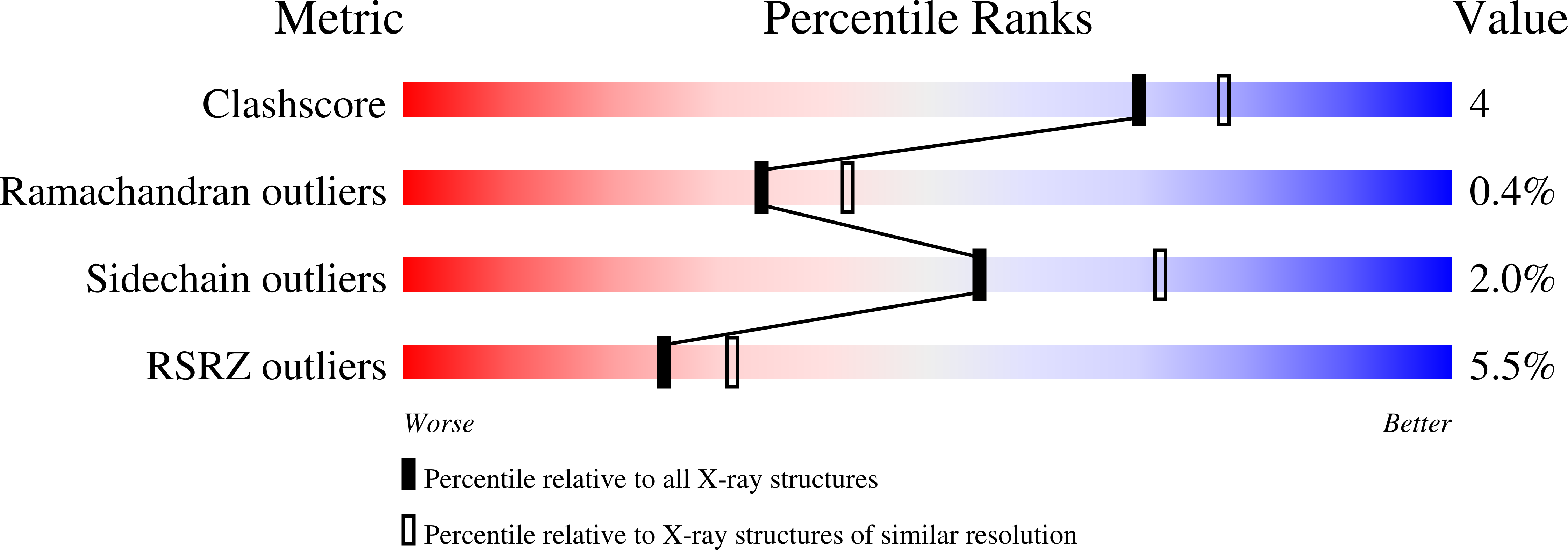
wwPDB Validation   3D Report Full Report


Ligand Structure Quality Assessment 


This is version 1.4 of the entry. See complete history


Literature

Crystal structure of the adenylyl cyclase activator Gsalpha

Sunahara, R.K.Tesmer, J.J.Gilman, A.G.Sprang, S.R.

(1997) Science 278: 1943-1947

  • DOI: https://doi.org/10.1126/science.278.5345.1943
  • Primary Citation of Related Structures:  
    1AZT

  • PubMed Abstract: 

    The crystal structure of Gsalpha, the heterotrimeric G protein alpha subunit that stimulates adenylyl cyclase, was determined at 2.5 A in a complex with guanosine 5'-O-(3-thiotriphosphate) (GTPgammaS). Gsalpha is the prototypic member of a family of GTP-binding proteins that regulate the activities of effectors in a hormone-dependent manner. Comparison of the structure of Gsalpha.GTPgammaS with that of Gialpha.GTPgammaS suggests that their effector specificity is primarily dictated by the shape of the binding surface formed by the switch II helix and the alpha3-beta5 loop, despite the high sequence homology of these elements. In contrast, sequence divergence explains the inability of regulators of G protein signaling to stimulate the GTPase activity of Gsalpha. The betagamma binding surface of Gsalpha is largely conserved in sequence and structure to that of Gialpha, whereas differences in the surface formed by the carboxyl-terminal helix and the alpha4-beta6 loop may mediate receptor specificity.


  • Organizational Affiliation

    Department of Pharmacology, The University of Texas Southwestern Medical Center, 5323 Harry Hines Boulevard, Dallas, TX 75235-9041, USA.


Macromolecules
Find similar proteins by:  (by identity cutoff)  |  3D Structure
Entity ID: 1
MoleculeChains Sequence LengthOrganismDetailsImage
GS-ALPHA
A, B
402Bos taurusMutation(s): 0 
Gene Names: GNAS
UniProt
Find proteins for P04896 (Bos taurus)
Explore P04896 
Go to UniProtKB:  P04896
Entity Groups  
Sequence Clusters30% Identity50% Identity70% Identity90% Identity95% Identity100% Identity
UniProt GroupP04896
Sequence Annotations
Expand
  • Reference Sequence
Small Molecules
Ligands 3 Unique
IDChains Name / Formula / InChI Key2D Diagram3D Interactions
GSP
Query on GSP

Download Ideal Coordinates CCD File 
M [auth A],
V [auth B]
5'-GUANOSINE-DIPHOSPHATE-MONOTHIOPHOSPHATE
C10 H16 N5 O13 P3 S
XOFLBQFBSOEHOG-UUOKFMHZSA-N
PO4
Query on PO4

Download Ideal Coordinates CCD File 
D [auth A]
E [auth A]
F [auth A]
G [auth A]
H [auth A]
D [auth A],
E [auth A],
F [auth A],
G [auth A],
H [auth A],
I [auth A],
J [auth A],
K [auth A],
L [auth A],
N [auth B],
O [auth B],
P [auth B],
R [auth B],
S [auth B],
T [auth B],
U [auth B]
PHOSPHATE ION
O4 P
NBIIXXVUZAFLBC-UHFFFAOYSA-K
MG
Query on MG

Download Ideal Coordinates CCD File 
C [auth A],
Q [auth B]
MAGNESIUM ION
Mg
JLVVSXFLKOJNIY-UHFFFAOYSA-N
Experimental Data & Validation

Experimental Data

  • Method: X-RAY DIFFRACTION
  • Resolution: 2.30 Å
  • R-Value Free: 0.282 
  • R-Value Work: 0.219 
  • R-Value Observed: 0.219 
  • Space Group: P 21 21 21
Unit Cell:
Length ( Å )Angle ( ˚ )
a = 88.3α = 90
b = 96.5β = 90
c = 133.4γ = 90
Software Package:
Software NamePurpose
AMoREphasing
X-PLORrefinement
DENZOdata reduction
SCALEPACKdata scaling

Structure Validation

View Full Validation Report


Ligand Structure Quality Assessment 


Entry History 

Deposition Data

Revision History  (Full details and data files)

  • Version 1.0: 1998-02-25
    Type: Initial release
  • Version 1.1: 2008-03-03
    Changes: Version format compliance
  • Version 1.2: 2011-07-13
    Changes: Version format compliance
  • Version 1.3: 2023-08-02
    Changes: Database references, Derived calculations, Refinement description
  • Version 1.4: 2024-05-22
    Changes: Data collection