1BXW | pdb_00001bxw

OUTER MEMBRANE PROTEIN A (OMPA) TRANSMEMBRANE DOMAIN


Experimental Data Snapshot

  • Method: X-RAY DIFFRACTION
  • Resolution: 2.50 Å
  • R-Value Free: 
    0.235 (Depositor) 
  • R-Value Work: 
    0.189 (Depositor), 0.190 (DCC) 

wwPDB Validation   3D Report Full Report


Ligand Structure Quality Assessment 

Created with Raphaël 2.3.0Worse 01 BetterLigand structure goodness of fit to experimental dataBest fitted C8EClick on this verticalbar to view details

This is version 1.6 of the entry. See complete history


Literature

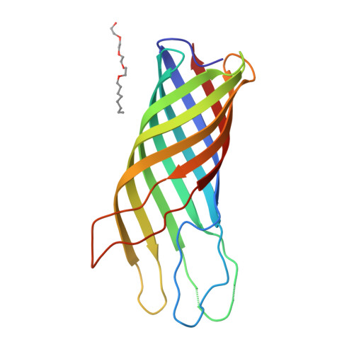
Structure of the outer membrane protein A transmembrane domain.

Pautsch, A.Schulz, G.E.

(1998) Nat Struct Biol 5: 1013-1017

  • DOI: https://doi.org/10.1038/2983
  • Primary Citation of Related Structures:  
    1BXW

  • PubMed Abstract: 

    The outer membrane protein A of Escherichia coli (OmpA) is an intensely studied example in the field of membrane protein folding. We have determined the structure of the OmpA transmembrane domain consisting of residues 1-171, by X-ray diffraction analysis, to a resolution of 2.5 A. It consists of a regular, extended eight-stranded beta-barrel and appears to be constructed like an inverse micelle with large water-filled cavities, but does not form a pore. Surprisingly, the cavities seem to be highly conserved during evolution. The structure corroborates the concept that all outer membrane proteins consist of beta-barrels. The structure constitutes a beta-barrel membrane anchor that appears to be the outer membrane equivalent of the single-chain alpha-helix anchor of the inner membrane.


  • Organizational Affiliation

    Institut für Organische Chemie und Biochemie, Albert-Ludwigs-Universität, Freiburg im Breisgau, Germany.


Macromolecules
Find similar proteins by:  (by identity cutoff)  |  3D Structure
Entity ID: 1
MoleculeChains Sequence LengthOrganismDetailsImage
PROTEIN (OUTER MEMBRANE PROTEIN A)172Escherichia coli BL21(DE3)Mutation(s): 3 
Gene Names: OMPA
Membrane Entity: Yes 
UniProt
Find proteins for P0A910 (Escherichia coli (strain K12))
Explore P0A910 
Go to UniProtKB:  P0A910
Entity Groups  
Sequence Clusters30% Identity50% Identity70% Identity90% Identity95% Identity100% Identity
UniProt GroupP0A910
Sequence Annotations
Expand
  • Reference Sequence
Small Molecules
Ligands 1 Unique
IDChains Name / Formula / InChI Key2D Diagram3D Interactions
C8E
Query on C8E

Download Ideal Coordinates CCD File 
B [auth A](HYDROXYETHYLOXY)TRI(ETHYLOXY)OCTANE
C16 H34 O5
FEOZZFHAVXYAMB-UHFFFAOYSA-N
Experimental Data & Validation

Experimental Data

  • Method: X-RAY DIFFRACTION
  • Resolution: 2.50 Å
  • R-Value Free:  0.235 (Depositor) 
  • R-Value Work:  0.189 (Depositor), 0.190 (DCC) 
Space Group: C 1 2 1
Unit Cell:
Length ( Å )Angle ( ˚ )
a = 69.18α = 90
b = 77.95β = 91.52
c = 50.93γ = 90
Software Package:
Software NamePurpose
SHARPphasing
REFMACrefinement
XDSdata reduction
CCP4data scaling

Structure Validation

View Full Validation Report


Ligand Structure Quality Assessment 

Created with Raphaël 2.3.0Worse 01 BetterLigand structure goodness of fit to experimental dataBest fitted C8EClick on this verticalbar to view details

Entry History 

Deposition Data

Revision History  (Full details and data files)

  • Version 1.0: 1998-10-14
    Type: Initial release
  • Version 1.1: 2008-04-27
    Changes: Version format compliance
  • Version 1.2: 2011-07-13
    Changes: Version format compliance
  • Version 1.3: 2017-11-29
    Changes: Advisory
  • Version 1.4: 2018-03-14
    Changes: Database references
  • Version 1.5: 2023-02-15
    Changes: Advisory, Database references, Derived calculations
  • Version 1.6: 2024-05-22
    Changes: Data collection