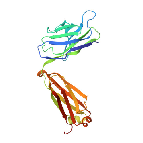
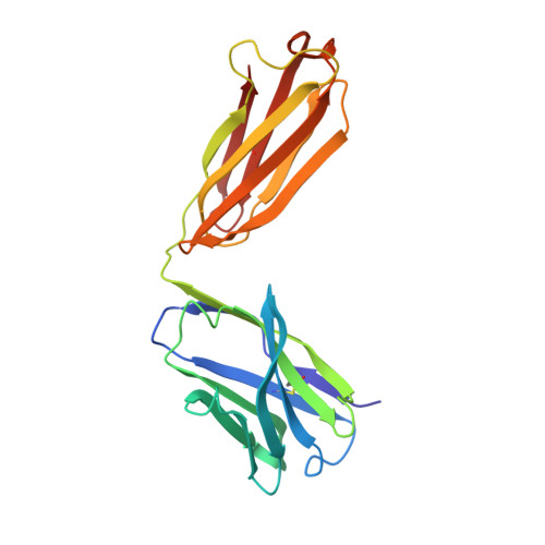
Local and transmitted conformational changes on complexation of an anti-sweetener Fab.
Guddat, L.W., Shan, L., Anchin, J.M., Linthicum, D.S., Edmundson, A.B.(1994) J Mol Biol 236: 247-274
- PubMed: 7893280
- DOI: https://doi.org/10.1006/jmbi.1994.1133
- Primary Citation of Related Structures:
1CGS, 2CGR - PubMed Abstract:
Crystal structures of an Fab (NC6.8) from a murine IgG2b(kappa) antibody and its complex with a sweet-tasting, N-,N'-,N"-trisubstituted guanidine compound (NC174) have been determined by X-ray analysis. Both crystal forms are produced by a microseeding technique in polyethylene glycol (PEG) 8000 but the habits and space groups are very different. The native protein crystallizes as plates in the monoclinic space group C2 and the complex crystallizes as prisms in the orthorhombic space group P2(1)2(1)2. The structures were solved by molecular replacement methods, with the Fab fragments from the 4-4-20, HyHel-5 and BV04-01 antibodies as starting models. On binding of the ligand, N-(p-cyanophenyl)-N'-(diphenylmethyl)-N"-(carboxymethyl)g uan idine, the protein exhibits significant local conformational changes in the active site, particularly in the third complementarity-determining region (CDR3) of the heavy chain. The ligand enters the small crevice by end-on insertion with the cyanophenyl group in the lead and the diphenyl rings partially protruding from the entrance. No strict pi-pi stacking interactions are observed. However, tyrosine L32 (CDR1), tyrosine L96 (CDR3) and tryptophan H33 (CDR1) help immobilize the cyanophenyl ring and guanido group, and tyrosine H96 moves about 4.5 A to lie between the rings of the diphenyl group. The positive charge on the guanido group is compensated by glutamic acid H50 (CDR2) while the negative charge on acetic acid is neutralized by arginine H56 (CDR2) and by hydrogen bonding with asparagine H58 (CDR2). Water molecules participate in the binding process by hydrogen bonding with the cyano and guanido groups. The mechanism of binding is a clear example of induced fit. Like hemoglobin, the NC6.8 Fab can be classified as an allosteric protein, since its overall structure is altered by the binding of a small ligand. In crystals of the native Fab the elbow bend angle is 184 degrees while in crystals of the complex the elbow angle is 153 degrees. There is also a reciprocal push-pull type of change where the heavy chain is flexed and the light chain is extended. The tail of the heavy chain, which would be connected to the Fc in an intact antibody, is displaced 19 A relative to its position in the unliganded Fab. Within the limited series of sweetener-Fab complexes we have thus far examined, only the NC174 hapten has produced such results.(ABSTRACT TRUNCATED AT 400 WORDS)
Organizational Affiliation:
Harrington Cancer Center, Amarillo, TX 79106.


