1DQD

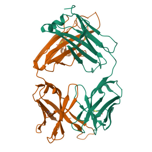
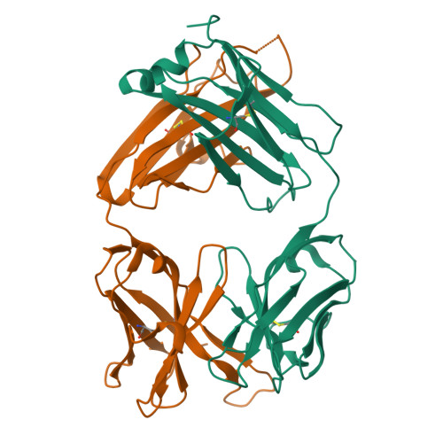
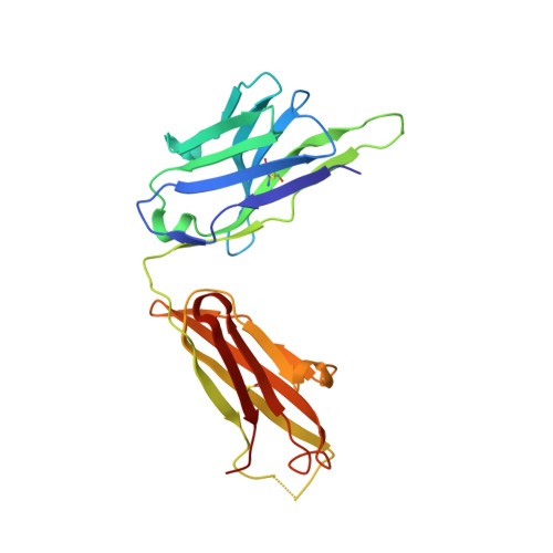
CRYSTAL STRUCTURE OF FAB HGR-2 F6, A COMPETITIVE ANTAGONIST OF THE GLUCAGON RECEPTOR


Experimental Data Snapshot

  • Method: X-RAY DIFFRACTION
  • Resolution: 2.10 Å
  • R-Value Free: 0.315 
  • R-Value Work: 0.217 

wwPDB Validation   3D Report Full Report


This is version 1.3 of the entry. See complete history


Literature

Structure of Fab hGR-2 F6, a competitive antagonist of the glucagon receptor.

Wright, L.M.Brzozowski, A.M.Hubbard, R.E.Pike, A.C.Roberts, S.M.Skovgaard, R.N.Svendsen, I.Vissing, H.Bywater, R.P.

(2000) Acta Crystallogr D Biol Crystallogr 56: 573-580

  • DOI: https://doi.org/10.1107/s090744490000233x
  • Primary Citation of Related Structures:  
    1DQD

  • PubMed Abstract: 

    The monoclonal antibody hGR-2 F6 has been raised against the human glucagon receptor and shown to act as a competitive antagonist. As a first step in the structural characterization of the receptor, the crystal structure of the Fab fragment from this antibody is reported at 2.1 A resolution. The hGR-2 F6 Fab crystallizes in the orthorhombic space group P2(1)2(1)2, with unit-cell parameters a = 76.14, b = 133.74, c = 37.46 A. A model generated by homology modelling was used as an aid in the chain-tracing and the Fab fragment structure was subsequently refined (final R factor = 21.7%). The structure obtained exhibits the typical immunoglobulin fold. Complementarity-determining regions (CDRs) L1, L2, L3, H1 and H2 could be superposed onto standard canonical CDR loops. The H3 loop could be classified according to recently published rules regarding loop length, sequence and conformation. This loop is 14 residues long, with an approximate beta-hairpin geometry, which is distorted somewhat by the presence of two trans proline residues at the beginning of the loop. It is expected that this H3 loop will facilitate the design of synthetic probes for the glucagon receptor that may be used to investigate receptor activity.


  • Organizational Affiliation

    York Structural Biology Laboratory, Department of Chemistry, University of York, York YO10 5DD, England.


Macromolecules
Find similar proteins by:  (by identity cutoff)  |  3D Structure
Entity ID: 1
MoleculeChains Sequence LengthOrganismDetailsImage
FAB HGR-2 F6A [auth L]221Mus musculusMutation(s): 0 
Entity Groups  
Sequence Clusters30% Identity50% Identity70% Identity90% Identity95% Identity100% Identity
Sequence Annotations
Expand
  • Reference Sequence
Find similar proteins by:  (by identity cutoff)  |  3D Structure
Entity ID: 2
MoleculeChains Sequence LengthOrganismDetailsImage
FAB HGR-2 F6B [auth H]222Mus musculusMutation(s): 0 
Entity Groups  
Sequence Clusters30% Identity50% Identity70% Identity90% Identity95% Identity100% Identity
Sequence Annotations
Expand
  • Reference Sequence
Experimental Data & Validation

Experimental Data

  • Method: X-RAY DIFFRACTION
  • Resolution: 2.10 Å
  • R-Value Free: 0.315 
  • R-Value Work: 0.217 
  • Space Group: P 21 21 2
Unit Cell:
Length ( Å )Angle ( ˚ )
a = 76.14α = 90
b = 133.74β = 90
c = 37.46γ = 90
Software Package:
Software NamePurpose
DENZOdata reduction
SCALEPACKdata scaling
AMoREphasing
REFMACrefinement

Structure Validation

View Full Validation Report


Entry History 

Deposition Data

Revision History  (Full details and data files)

  • Version 1.0: 2000-05-10
    Type: Initial release
  • Version 1.1: 2008-04-27
    Changes: Version format compliance
  • Version 1.2: 2011-07-13
    Changes: Version format compliance
  • Version 1.3: 2024-10-30
    Changes: Data collection, Database references, Structure summary