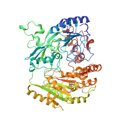
1E0Y

Structure of the D170S/T457E double mutant of vanillyl-alcohol oxidase


Experimental Data Snapshot

  • Method: X-RAY DIFFRACTION
  • Resolution: 2.75 Å
  • R-Value Free: 0.287 
  • R-Value Work: 0.225 
  • R-Value Observed: 0.225 

Starting Model: experimental
View more details

wwPDB Validation   3D Report Full Report


Ligand Structure Quality Assessment 


This is version 1.4 of the entry. See complete history


Literature

Inversion of Stereospecificity in Vanillyl-Alcohol Oxidase

Van Der Heuvel, R.H.H.Fraaije, M.W.Espinosa, M.F.Mattevi, A.Van Berkel, W.J.H.

(2000) Proc Natl Acad Sci U S A 97: 9455

  • DOI: https://doi.org/10.1073/pnas.160175897
  • Primary Citation of Related Structures:  
    1E0Y

  • PubMed Abstract: 

    Vanillyl-alcohol oxidase (VAO) is the prototype of a newly recognized family of structurally related oxidoreductases sharing a conserved FAD-binding domain. The active site of VAO is formed by a cavity where the enzyme is able to catalyze many reactions with phenolic substrates. Among these reactions is the stereospecific hydroxylation of 4-ethylphenol-forming (R)-1-(4'-hydroxyphenyl)ethanol. During this conversion, Asp-170 is probably critical for the hydration of the initially formed p-quinone methide intermediate. By site-directed mutagenesis, the putative active site base has been relocated to the opposite face of the active site cavity. In this way, a change in stereospecificity has been achieved. Like native VAO, the single mutants T457E, D170A, and D170S preferentially converted 4-ethylphenol to the (R)-enantiomer of 1-(4'-hydroxyphenyl)ethanol. The double mutants D170A/T457E and D170S/T457E exhibited an inverted stereospecificity with 4-ethylphenol. Particularly, D170S/T457E was strongly (S)-selective, with an enantiomeric excess of 80%. The crystal structure of D170S/T457E, in complex with trifluoromethylphenol, showed a highly conserved mode of ligand binding and revealed that the distinctive catalytic properties of this mutant are not caused by major structural changes.


  • Organizational Affiliation

    Department of Biomolecular Sciences, Laboratory of Biochemistry, Wageningen University, The Netherlands.


Macromolecules
Find similar proteins by:  (by identity cutoff)  |  3D Structure
Entity ID: 1
MoleculeChains Sequence LengthOrganismDetailsImage
VANILLYL-ALCOHOL OXIDASE
A, B
560Penicillium simplicissimumMutation(s): 2 
EC: 1.1.3.38
UniProt
Find proteins for P56216 (Penicillium simplicissimum)
Explore P56216 
Go to UniProtKB:  P56216
Entity Groups  
Sequence Clusters30% Identity50% Identity70% Identity90% Identity95% Identity100% Identity
UniProt GroupP56216
Sequence Annotations
Expand
  • Reference Sequence
Small Molecules
Ligands 2 Unique
IDChains Name / Formula / InChI Key2D Diagram3D Interactions
FAD
Query on FAD

Download Ideal Coordinates CCD File 
C [auth A],
D [auth B]
FLAVIN-ADENINE DINUCLEOTIDE
C27 H33 N9 O15 P2
VWWQXMAJTJZDQX-UYBVJOGSSA-N
FCR
Query on FCR

Download Ideal Coordinates CCD File 
E [auth B]ALPHA,ALPHA,ALPHA-TRIFLUORO-P-CRESOL
C7 H5 F3 O
BAYGVMXZJBFEMB-UHFFFAOYSA-N
Experimental Data & Validation

Experimental Data

  • Method: X-RAY DIFFRACTION
  • Resolution: 2.75 Å
  • R-Value Free: 0.287 
  • R-Value Work: 0.225 
  • R-Value Observed: 0.225 
  • Space Group: I 4
Unit Cell:
Length ( Å )Angle ( ˚ )
a = 131.281α = 90
b = 131.281β = 90
c = 134.355γ = 90
Software Package:
Software NamePurpose
REFMACrefinement
MOSFLMdata reduction
CCP4data scaling
CCP4phasing

Structure Validation

View Full Validation Report


Ligand Structure Quality Assessment 


Entry History 

Deposition Data

Revision History  (Full details and data files)

  • Version 1.0: 2000-04-12
    Type: Initial release
  • Version 1.1: 2011-05-08
    Changes: Version format compliance
  • Version 1.2: 2011-07-13
    Changes: Version format compliance
  • Version 1.3: 2019-07-24
    Changes: Advisory, Data collection, Derived calculations
  • Version 1.4: 2023-12-06
    Changes: Advisory, Data collection, Database references, Derived calculations, Other, Refinement description