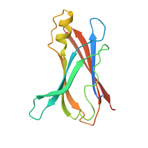
1E5A

Structure of human transthyretin complexed with bromophenols: a new mode of binding


Experimental Data Snapshot

  • Method: X-RAY DIFFRACTION
  • Resolution: 1.80 Å
  • R-Value Free: 0.262 
  • R-Value Work: 0.214 
  • R-Value Observed: 0.214 

Starting Model: experimental
View more details

wwPDB Validation   3D Report Full Report


This is version 1.3 of the entry. See complete history


Literature

Structure of Human Transthyretin Complexed with Bromophenols : A New Mode of Binding

Ghosh, M.Meerts, I.A.T.M.Cook, A.Bergman, A.Brouwer, A.Johnson, L.N.

(2000) Acta Crystallogr D Biol Crystallogr 56: 1085

  • DOI: https://doi.org/10.1107/s0907444900008568
  • Primary Citation of Related Structures:  
    1E3F, 1E4H, 1E5A

  • PubMed Abstract: 

    The binding of two organohalogen substances, pentabromophenol (PBP) and 2,4,6-tribromophenol (TBP), to human transthyretin (TTR), a thyroid hormone transport protein, has been studied by in vitro competitive binding assays and by X-ray crystallography. Both compounds bind to TTR with high affinity, in competition with the natural ligand thyroxine (T(4)). The crystal structures of the TTR-PBP and TTR-TBP complexes show some unusual binding patterns for the ligands. They bind exclusively in the 'reversed' mode, with their hydroxyl group pointing towards the mouth of the binding channel and in planes approximately perpendicular to that adopted by the T(4) phenolic ring in a TTR-T(4) complex, a feature not observed before. The hydroxyl group in the ligands, which was previously thought to be a key ingredient for a strong binding to TTR, does not seem to play an important role in the binding of these compounds to TTR. In the TTR-PBP complex, it is primarily the halogens which interact with the TTR molecule and therefore must account for the strong affinity of binding. The interactions with the halogens are smaller in number in TTR-TBP and there is a decrease in affinity, even though the interaction with the hydroxyl group is stronger than that in the TTR-PBP complex.


  • Organizational Affiliation

    Laboratory of Molecular Biophysics, Department of Biochemistry, South Parks Road, Oxford OX1 3QU, England. mina@biop.ox.ac.uk


Macromolecules
Find similar proteins by:  (by identity cutoff)  |  3D Structure
Entity ID: 1
MoleculeChains Sequence LengthOrganismDetailsImage
TRANSTHYRETIN
A, B
127Homo sapiensMutation(s): 0 
UniProt & NIH Common Fund Data Resources
Find proteins for P02766 (Homo sapiens)
Go to UniProtKB:  P02766
PHAROS:  P02766
GTEx:  ENSG00000118271 
Sequence Annotations
Expand
  • Reference Sequence
Small Molecules
Binding Affinity Annotations 
IDSourceBinding Affinity
TBP PDBBind:  1E5A Kd: 23 (nM) from 1 assay(s)
Experimental Data & Validation

Experimental Data

  • Method: X-RAY DIFFRACTION
  • Resolution: 1.80 Å
  • R-Value Free: 0.262 
  • R-Value Work: 0.214 
  • R-Value Observed: 0.214 
  • Space Group: P 21 21 2
Unit Cell:
Length ( Å )Angle ( ˚ )
a = 42.8α = 90
b = 84.9β = 90
c = 64.4γ = 90
Software Package:
Software NamePurpose
X-PLORrefinement
DENZOdata reduction
SCALEPACKdata scaling
X-PLORphasing

Structure Validation

View Full Validation Report


Entry History 

Deposition Data

Revision History  (Full details and data files)

  • Version 1.0: 2000-08-29
    Type: Initial release
  • Version 1.1: 2011-08-10
    Changes: Atomic model, Data collection, Database references, Derived calculations, Non-polymer description, Other, Structure summary, Version format compliance
  • Version 1.2: 2017-07-05
    Changes: Data collection
  • Version 1.3: 2023-12-13
    Changes: Data collection, Database references, Derived calculations, Refinement description