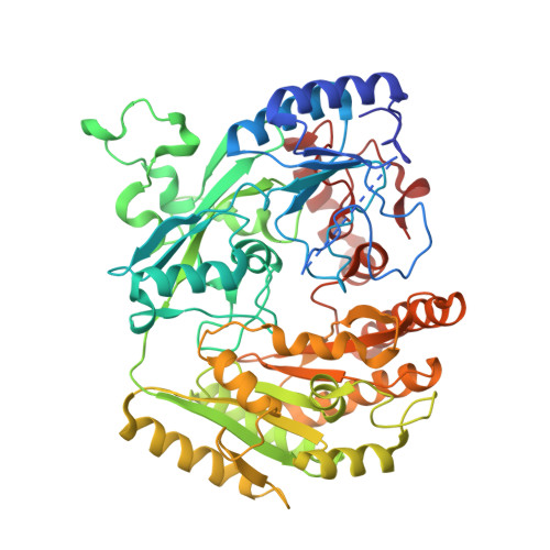
1E8F

STRUCTURE OF THE H61T MUTANT OF THE FLAVOENZYME VANILLYL-ALCOHOL OXIDASE IN THE APO FORM


Experimental Data Snapshot

  • Method: X-RAY DIFFRACTION
  • Resolution: 2.90 Å
  • R-Value Free: 0.312 
  • R-Value Work: 0.239 
  • R-Value Observed: 0.239 

Starting Model: experimental
View more details

wwPDB Validation   3D Report Full Report


This is version 1.3 of the entry. See complete history


Literature

Structural Analysis of Flavinylation in Vanillyl-Alcohol Oxidase

Fraaije, M.W.Van Der Heuvel, R.H.H.Van Berkel, W.J.H.Mattevi, A.

(2001) J Biol Chem 275: 38654

  • DOI: https://doi.org/10.1074/jbc.M004753200
  • Primary Citation of Related Structures:  
    1E8F, 1E8G, 1E8H

  • PubMed Abstract: 

    Vanillyl-alcohol oxidase (VAO) is member of a newly recognized flavoprotein family of structurally related oxidoreductases. The enzyme contains a covalently linked FAD cofactor. To study the mechanism of flavinylation we have created a design point mutation (His-61 --> Thr). In the mutant enzyme the covalent His-C8alpha-flavin linkage is not formed, while the enzyme is still able to bind FAD and perform catalysis. The H61T mutant displays a similar affinity for FAD and ADP (K(d) = 1.8 and 2.1 microm, respectively) but does not interact with FMN. H61T is about 10-fold less active with 4-(methoxymethyl)phenol) (k(cat) = 0.24 s(-)(1), K(m) = 40 microm) than the wild-type enzyme. The crystal structures of both the holo and apo form of H61T are highly similar to the structure of wild-type VAO, indicating that binding of FAD to the apoprotein does not require major structural rearrangements. These results show that covalent flavinylation is an autocatalytical process in which His-61 plays a crucial role by activating His-422. Furthermore, our studies clearly demonstrate that in VAO, the FAD binds via a typical lock-and-key approach to a preorganized binding site.


  • Organizational Affiliation

    Department of Biomolecular Sciences, Laboratory of Biochemistry, Wageningen University, Dreijenlaan 3, 6703 HA Wageningen, The Netherlands. m.w.fraaije@chem.rug.nl


Macromolecules
Find similar proteins by:  (by identity cutoff)  |  3D Structure
Entity ID: 1
MoleculeChains Sequence LengthOrganismDetailsImage
VANILLYL-ALCOHOL OXIDASE
A, B
560Penicillium simplicissimumMutation(s): 1 
EC: 1.1.3.7 (PDB Primary Data), 1.1.3.38 (UniProt)
UniProt
Find proteins for P56216 (Penicillium simplicissimum)
Explore P56216 
Go to UniProtKB:  P56216
Entity Groups  
Sequence Clusters30% Identity50% Identity70% Identity90% Identity95% Identity100% Identity
UniProt GroupP56216
Sequence Annotations
Expand
  • Reference Sequence
Experimental Data & Validation

Experimental Data

  • Method: X-RAY DIFFRACTION
  • Resolution: 2.90 Å
  • R-Value Free: 0.312 
  • R-Value Work: 0.239 
  • R-Value Observed: 0.239 
  • Space Group: I 4
Unit Cell:
Length ( Å )Angle ( ˚ )
a = 129.28α = 90
b = 129.28β = 90
c = 130.4γ = 90
Software Package:
Software NamePurpose
REFMACrefinement
MOSFLMdata reduction
SCALAdata scaling
CCP4phasing

Structure Validation

View Full Validation Report


Entry History 

Deposition Data

Revision History  (Full details and data files)

  • Version 1.0: 2000-09-21
    Type: Initial release
  • Version 1.1: 2011-05-08
    Changes: Version format compliance
  • Version 1.2: 2011-07-13
    Changes: Version format compliance
  • Version 1.3: 2023-12-13
    Changes: Data collection, Database references, Other, Refinement description