Crystal structure of unligated guanylate kinase from yeast reveals GMP-induced conformational changes.
Blaszczyk, J., Li, Y., Yan, H., Ji, X.(2001) J Mol Biol 307: 247-257
- PubMed: 11243817
- DOI: https://doi.org/10.1006/jmbi.2000.4427
- Primary Citation of Related Structures:
1EX6, 1EX7 - PubMed Abstract:
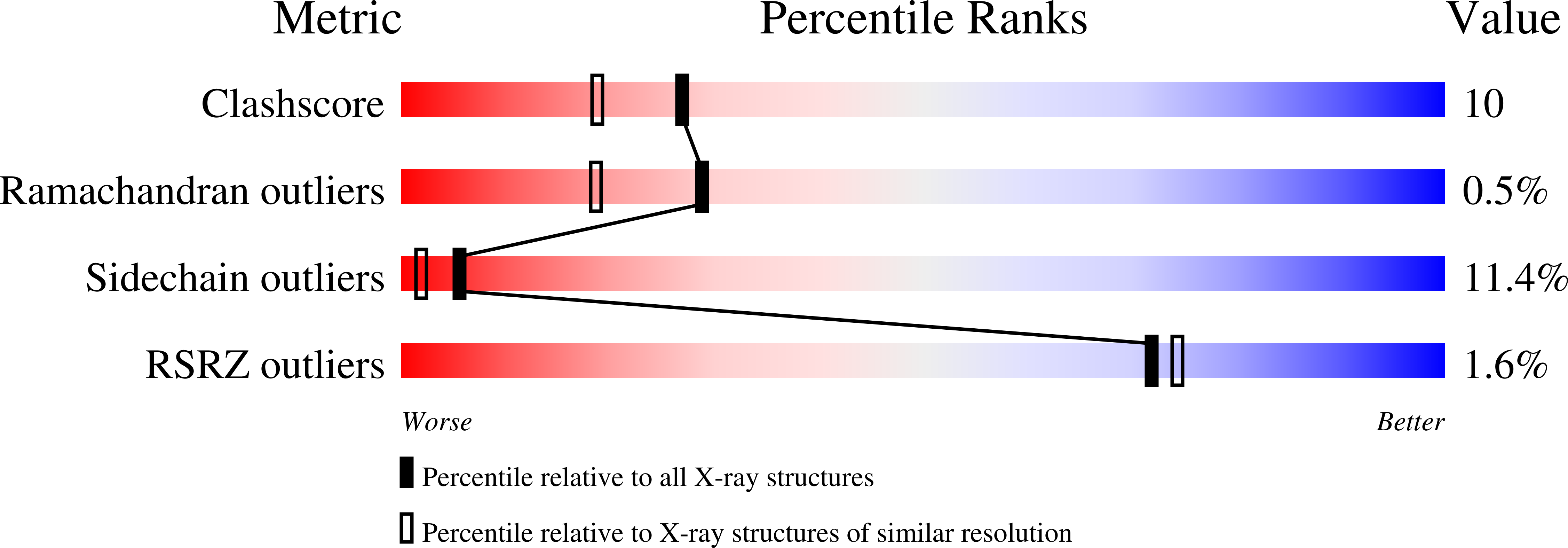
The crystal structure of guanylate kinase (GK) from yeast (Saccharomyces cerevisiae) with a non-acetylated N terminus has been determined in its unligated form (apo-GK) as well as in complex with GMP (GK.GMP). The structure of apo-GK was solved with multiwavelength anomalous diffraction data and refined to an R-factor of 0.164 (R(free)=0.199) at 2.3 A resolution. The structure of GK.GMP was determined using the crystal structure of GK with an acetylated N terminus as the search model and refined to an R-factor of 0.156 (R(free)=0.245) at 1.9 A. GK belongs to the family of nucleoside monophosphate (NMP) kinases and catalyzes the reversible phosphoryl transfer from ATP to GMP. Like other NMP kinases, GK consists of three dynamic domains: the CORE, LID, and NMP-binding domains. Dramatic movements of the GMP-binding domain and smaller but significant movements of the LID domain have been revealed by comparing the structures of apo-GK and GK.GMP. apo-GK has a much more open conformation than the GK.GMP complex. Systematic analysis of the domain movements using the program DynDom shows that the large movements of the GMP-binding domain involve a rotation around an effective hinge axis approximately parallel with helix 3, which connects the GMP-binding and CORE domains. The C-terminal portion of helix 3, which connects to the CORE domain, has strikingly higher temperature factors in GK.GMP than in apo-GK, indicating that these residues become more mobile upon GMP binding. The results suggest that helix 3 plays an important role in domain movement. Unlike the GMP-binding domain, which moves toward the active center of the enzyme upon GMP binding, the LID domain moves away from the active center and makes the presumed ATP-binding site more open. Therefore, the LID domain movement may facilitate the binding of MgATP. The structure of the recombinant GK.GMP complex superimposes very well with that of the native GK.GMP complex, indicating that N-terminal acetylation does not have significant impact on the three-dimensional structure of GK.
Organizational Affiliation:
National Cancer Institute, Macromolecular Crystallography Laboratory, Frederick, MD 21702, USA.


