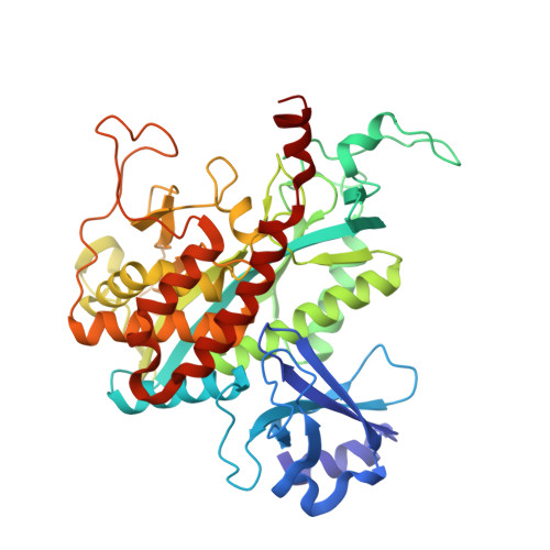
1FPY

CRYSTAL STRUCTURE OF GLUTAMINE SYNTHETASE FROM SALMONELLA TYPHIMURIUM WITH INHIBITOR PHOSPHINOTHRICIN


Experimental Data Snapshot

  • Method: X-RAY DIFFRACTION
  • Resolution: 2.89 Å
  • R-Value Free: 0.263 
  • R-Value Work: 0.248 
  • R-Value Observed: 0.250 

wwPDB Validation   3D Report Full Report


Ligand Structure Quality Assessment 


This is version 1.3 of the entry. See complete history


Literature

The crystal structure of phosphinothricin in the active site of glutamine synthetase illuminates the mechanism of enzymatic inhibition.

Gill, H.S.Eisenberg, D.

(2001) Biochemistry 40: 1903-1912

  • DOI: https://doi.org/10.1021/bi002438h
  • Primary Citation of Related Structures:  
    1F1H, 1F52, 1FPY

  • PubMed Abstract: 

    Phosphinothricin is a potent inhibitor of the enzyme glutamine synthetase (GS). The resolution of the native structure of GS from Salmonella typhimurium has been extended to 2.5 A resolution, and the improved model is used to determine the structure of phosphinothricin complexed to GS by difference Fourier methods. The structure suggests a noncovalent, dead-end mechanism of inhibition. Phosphinothricin occupies the glutamate substrate pocket and stabilizes the Glu327 flap in a position which blocks the glutamate entrance to the active site, trapping the inhibitor on the enzyme. One oxygen of the phosphinyl group of phosphinothricin appears to be protonated, because of its proximity to the carboxylate group of Glu327. The other phosphinyl oxygen protrudes into the negatively charged binding pocket for the substrate ammonium, disrupting that pocket. The distribution of charges in the glutamate binding pocket is complementary to those of phosphinothricin. The presence of a second ammonium binding site within the active site is confirmed by its analogue thallous ion, marking the ammonium site and its protein ligands. The inhibition of GS by methionine sulfoximine can be explained by the same mechanism. These models of inhibited GS further illuminate its catalytic mechanism.


  • Organizational Affiliation

    UCLA-DOE Lab of Structural Biology and Molecular Medicine, Box 951570, University of California, Los Angeles, California 90095-1570, USA.


Macromolecules
Find similar proteins by:  (by identity cutoff)  |  3D Structure
Entity ID: 1
MoleculeChains Sequence LengthOrganismDetailsImage
GLUTAMINE SYNTHETASE
A, B, C, D, E
A, B, C, D, E, F, G, H, I, J, K, L
468Salmonella enterica subsp. enterica serovar TyphimuriumMutation(s): 0 
EC: 6.3.1.2
UniProt
Find proteins for P0A1P6 (Salmonella typhimurium (strain LT2 / SGSC1412 / ATCC 700720))
Explore P0A1P6 
Go to UniProtKB:  P0A1P6
Entity Groups  
Sequence Clusters30% Identity50% Identity70% Identity90% Identity95% Identity100% Identity
UniProt GroupP0A1P6
Sequence Annotations
Expand
  • Reference Sequence
Small Molecules
Ligands 3 Unique
IDChains Name / Formula / InChI Key2D Diagram3D Interactions
ADP
Query on ADP

Download Ideal Coordinates CCD File 
AA [auth D]
CB [auth K]
EA [auth E]
GB [auth L]
IA [auth F]
AA [auth D],
CB [auth K],
EA [auth E],
GB [auth L],
IA [auth F],
MA [auth G],
O [auth A],
QA [auth H],
S [auth B],
UA [auth I],
W [auth C],
YA [auth J]
ADENOSINE-5'-DIPHOSPHATE
C10 H15 N5 O10 P2
XTWYTFMLZFPYCI-KQYNXXCUSA-N
PPQ
Query on PPQ

Download Ideal Coordinates CCD File 
BA [auth D]
DB [auth K]
FA [auth E]
HB [auth L]
JA [auth F]
BA [auth D],
DB [auth K],
FA [auth E],
HB [auth L],
JA [auth F],
NA [auth G],
P [auth A],
RA [auth H],
T [auth B],
VA [auth I],
X [auth C],
ZA [auth J]
PHOSPHINOTHRICIN
C5 H12 N O4 P
IAJOBQBIJHVGMQ-BYPYZUCNSA-N
MN
Query on MN

Download Ideal Coordinates CCD File 
AB [auth K]
BB [auth K]
CA [auth E]
DA [auth E]
EB [auth L]
AB [auth K],
BB [auth K],
CA [auth E],
DA [auth E],
EB [auth L],
FB [auth L],
GA [auth F],
HA [auth F],
KA [auth G],
LA [auth G],
M [auth A],
N [auth A],
OA [auth H],
PA [auth H],
Q [auth B],
R [auth B],
SA [auth I],
TA [auth I],
U [auth C],
V [auth C],
WA [auth J],
XA [auth J],
Y [auth D],
Z [auth D]
MANGANESE (II) ION
Mn
WAEMQWOKJMHJLA-UHFFFAOYSA-N
Binding Affinity Annotations 
IDSourceBinding Affinity
PPQ PDBBind:  1FPY Ki: 1000 (nM) from 1 assay(s)
Experimental Data & Validation

Experimental Data

  • Method: X-RAY DIFFRACTION
  • Resolution: 2.89 Å
  • R-Value Free: 0.263 
  • R-Value Work: 0.248 
  • R-Value Observed: 0.250 
  • Space Group: C 1 2 1
Unit Cell:
Length ( Å )Angle ( ˚ )
a = 230.6α = 90
b = 132.5β = 102.4
c = 195.9γ = 90
Software Package:
Software NamePurpose
X-PLORmodel building
X-PLORrefinement
DENZOdata reduction
SCALEPACKdata scaling
X-PLORphasing

Structure Validation

View Full Validation Report


Ligand Structure Quality Assessment 


Entry History 

Deposition Data

Revision History  (Full details and data files)

  • Version 1.0: 2001-04-04
    Type: Initial release
  • Version 1.1: 2008-04-27
    Changes: Version format compliance
  • Version 1.2: 2011-07-13
    Changes: Version format compliance
  • Version 1.3: 2024-02-07
    Changes: Data collection, Database references, Derived calculations