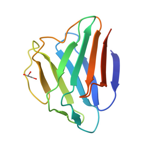
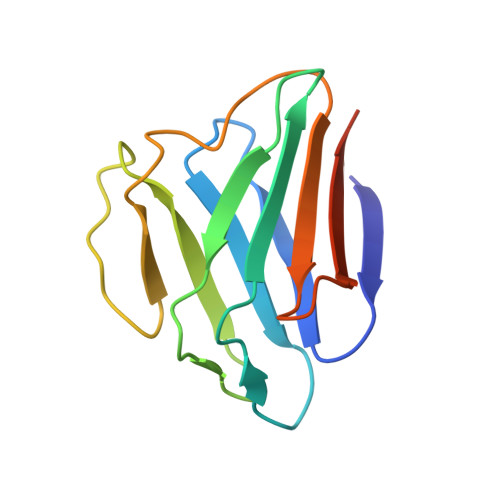
Structure of the globular tail of nuclear lamin.
Dhe-Paganon, S., Werner, E.D., Chi, Y.I., Shoelson, S.E.(2002) J Biological Chem 277: 17381-17384
- PubMed: 11901143
- DOI: https://doi.org/10.1074/jbc.C200038200
- Primary Citation of Related Structures:
1IFR - PubMed Abstract:
The nuclear lamins form a two-dimensional matrix that provides integrity to the cell nucleus and participates in nuclear activities. Mutations in the region of human LMNA encoding the carboxyl-terminal tail Lamin A/C are associated with forms of muscular dystrophy and familial partial lipodystrophy (FPLD). To help discriminate tissue-specific phenotypes, we have solved at 1.4-A resolution the three-dimensional crystal structure of the lamin A/C globular tail. The domain adopts a novel, all beta immunoglobulin-like fold. FPLD-associated mutations cluster within a small surface, whereas muscular dystrophy-associated mutations are distributed throughout the protein core and on its surface. These findings distinguish myopathy- and lipodystrophy-associated mutations and provide a structural framework for further testing hypotheses concerning lamin function.
Organizational Affiliation:
Joslin Diabetes Center & Department of Medicine, Harvard Medical School, Boston, Massachusetts 02215, USA.