1J9E

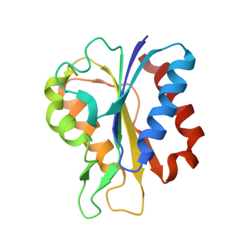
Low Temperature (100K) Crystal Structure of Flavodoxin D. vulgaris S35C Mutant at 1.44 Angstrom Resolution


Experimental Data Snapshot

  • Method: X-RAY DIFFRACTION
  • Resolution: 1.44 Å
  • R-Value Free: 0.156 
  • R-Value Work: 0.136 
  • R-Value Observed: 0.139 

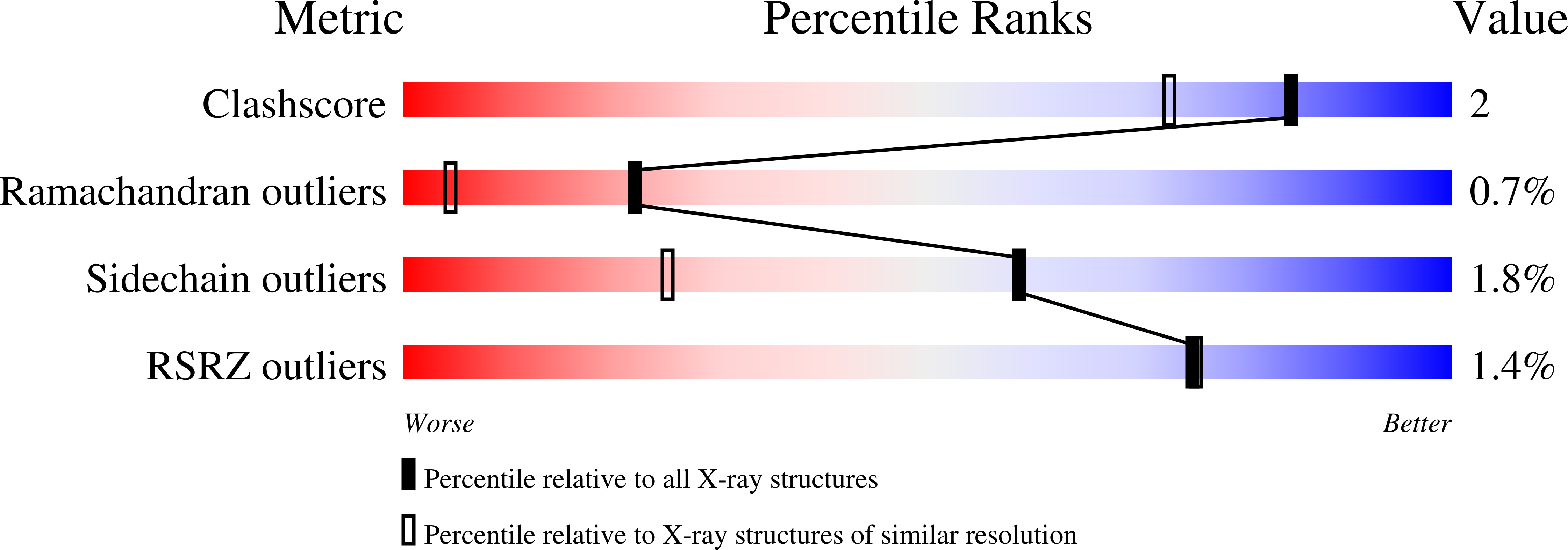
wwPDB Validation   3D Report Full Report


Ligand Structure Quality Assessment 


This is version 1.4 of the entry. See complete history


Literature

Comparison of the refined crystal structures of wild-type (1.34 A) flavodoxin from Desulfovibrio vulgaris and the S35C mutant (1.44 A) at 100 K.

Artali, R.Bombieri, G.Meneghetti, F.Gilardi, G.Sadeghi, S.J.Cavazzini, D.Rossi, G.L.

(2002) Acta Crystallogr D Biol Crystallogr 58: 1787-1792

  • DOI: https://doi.org/10.1107/s0907444902012234
  • Primary Citation of Related Structures:  
    1J8Q, 1J9E, 1J9G

  • PubMed Abstract: 

    Engineered flavodoxins in which a surface residue has been replaced by an exposed cysteine are useful modules to link multi-domain redox proteins obtained by gene fusion to electrode surfaces. In the present work, the crystal structure of the S35C mutant of Desulfovibrio vulgaris flavodoxin in the oxidized state has been determined and compared with a refined structure of the wild type (wt). The structure of wt flavodoxin (space group P4(3)2(1)2, unit-cell parameters a = 50.52, b = 50.52, c = 138.59 A) at 1.34 A resolution has been refined to R = 0.16 and R(free) = 0.18. The structure of the S35C mutant (space group P4(3)2(1)2, unit-cell parameters a = 50.55, b = 50.55, c = 138.39 A) at 1.44 A resolution has been refined to R = 0.13 and R(free) = 0.16. Data sets were collected with synchrotron radiation at 100 K. In the S35C mutant, the Cys35 thiol group points towards a hydrophobic region, whilst in the wt the Ser35 hydroxyl group points towards a more polar region. The solvent exposure of Cys35 is 43 A(2), of which 8 A(2) is for the sulfur. This is comparable to the exposure of 48 A(2) found for the wt Ser35, where that of the hydroxyl oxygen is also 8 A(2).


  • Organizational Affiliation

    Institute of Pharmaceutical Chemistry, University of Milano, Italy.


Macromolecules
Find similar proteins by:  (by identity cutoff)  |  3D Structure
Entity ID: 1
MoleculeChains Sequence LengthOrganismDetailsImage
FLAVODOXIN147Nitratidesulfovibrio vulgarisMutation(s): 1 
UniProt
Find proteins for P00323 (Nitratidesulfovibrio vulgaris (strain ATCC 29579 / DSM 644 / CCUG 34227 / NCIMB 8303 / VKM B-1760 / Hildenborough))
Explore P00323 
Go to UniProtKB:  P00323
Entity Groups  
Sequence Clusters30% Identity50% Identity70% Identity90% Identity95% Identity100% Identity
UniProt GroupP00323
Sequence Annotations
Expand
  • Reference Sequence
Small Molecules
Ligands 1 Unique
IDChains Name / Formula / InChI Key2D Diagram3D Interactions
FMN
Query on FMN

Download Ideal Coordinates CCD File 
B [auth A]FLAVIN MONONUCLEOTIDE
C17 H21 N4 O9 P
FVTCRASFADXXNN-SCRDCRAPSA-N
Experimental Data & Validation

Experimental Data

  • Method: X-RAY DIFFRACTION
  • Resolution: 1.44 Å
  • R-Value Free: 0.156 
  • R-Value Work: 0.136 
  • R-Value Observed: 0.139 
  • Space Group: P 43 21 2
Unit Cell:
Length ( Å )Angle ( ˚ )
a = 50.55α = 90
b = 50.55β = 90
c = 138.39γ = 90
Software Package:
Software NamePurpose
MOSFLMdata reduction
SCALAdata scaling
SHELXL-97model building
SHELXL-97refinement
CCP4data scaling
SHELXL-97phasing

Structure Validation

View Full Validation Report


Ligand Structure Quality Assessment 


Entry History 

Deposition Data

Revision History  (Full details and data files)

  • Version 1.0: 2001-09-05
    Type: Initial release
  • Version 1.1: 2008-04-27
    Changes: Version format compliance
  • Version 1.2: 2011-07-13
    Changes: Version format compliance
  • Version 1.3: 2021-10-27
    Changes: Database references, Derived calculations
  • Version 1.4: 2024-02-07
    Changes: Data collection