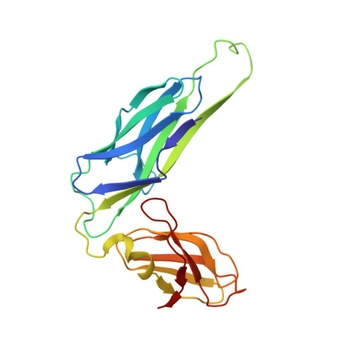
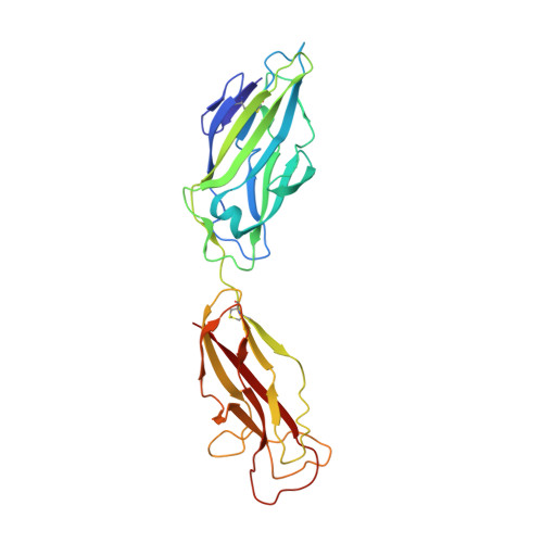
1KLF

FIMH ADHESIN-FIMC CHAPERONE COMPLEX WITH D-MANNOSE


Experimental Data Snapshot

  • Method: X-RAY DIFFRACTION
  • Resolution: 2.79 Å
  • R-Value Free: 0.280 
  • R-Value Work: 0.239 
  • R-Value Observed: 0.239 

Starting Model: experimental
View more details

wwPDB Validation   3D Report Full Report


Ligand Structure Quality Assessment 


This is version 1.5 of the entry. See complete history


Literature

Structural basis of tropism of Escherichia coli to the bladder during urinary tract infection.

Hung, C.S.Bouckaert, J.Hung, D.Pinkner, J.Widberg, C.DeFusco, A.Auguste, C.G.Strouse, R.Langermann, S.Waksman, G.Hultgren, S.J.

(2002) Mol Microbiol 44: 903-915

  • DOI: https://doi.org/10.1046/j.1365-2958.2002.02915.x
  • Primary Citation of Related Structures:  
    1KIU, 1KLF

  • PubMed Abstract: 

    The first step in the colonization of the human urinary tract by pathogenic Escherichia coli is the mannose-sensitive binding of FimH, the adhesin present at the tip of type 1 pili, to the bladder epithelium. We elucidated crystallographically the interactions of FimH with D-mannose. The unique site binding pocket occupied by D-mannose was probed using site-directed mutagenesis. All but one of the mutants examined had greatly diminished mannose-binding activity and had also lost the ability to bind human bladder cells. The binding activity of the mono-saccharide D-mannose was delineated from this of mannotriose (Man(alpha 1-3)[Man(alpha 1-6)]Man) by generating mutants that abolished D-mannose binding but retained mannotriose binding activity. Our structure/function analysis demonstrated that the binding of the monosaccharide alpha-D-mannose is the primary bladder cell receptor for uropathogenic E. coli and that this event requires a highly conserved FimH binding pocket. The residues in the FimH mannose-binding pocket were sequenced and found to be invariant in over 200 uropathogenic strains of E. coli. Only enterohaemorrhagic E. coli (EHEC) possess a sequence variation within the mannose-binding pocket of FimH, suggesting a naturally occurring mechanism of attenuation in EHEC bacteria that would prevent them from being targeted to the urinary tract.


  • Organizational Affiliation

    Department of Molecular Microbiology, Washington University School of Medicine, St. Louis, MO 63110, USA.


Macromolecules
Find similar proteins by:  (by identity cutoff)  |  3D Structure
Entity ID: 1
MoleculeChains Sequence LengthOrganismDetailsImage
CHAPERONE PROTEIN FIMC
A, C, E, G, I
A, C, E, G, I, K, M, O
205Escherichia coliMutation(s): 0 
Gene Names: FIMC
UniProt
Find proteins for P31697 (Escherichia coli (strain K12))
Explore P31697 
Go to UniProtKB:  P31697
Entity Groups  
Sequence Clusters30% Identity50% Identity70% Identity90% Identity95% Identity100% Identity
UniProt GroupP31697
Sequence Annotations
Expand
  • Reference Sequence
Find similar proteins by:  (by identity cutoff)  |  3D Structure
Entity ID: 2
MoleculeChains Sequence LengthOrganismDetailsImage
FIMH PROTEIN
B, D, F, H, J
B, D, F, H, J, L, N, P
279Escherichia coliMutation(s): 0 
Gene Names: FIMH
UniProt
Find proteins for P08191 (Escherichia coli (strain K12))
Explore P08191 
Go to UniProtKB:  P08191
Entity Groups  
Sequence Clusters30% Identity50% Identity70% Identity90% Identity95% Identity100% Identity
UniProt GroupP08191
Sequence Annotations
Expand
  • Reference Sequence
Small Molecules
Binding Affinity Annotations 
IDSourceBinding Affinity
MAN BindingDB:  1KLF Kd: 2300 (nM) from 1 assay(s)
Experimental Data & Validation

Experimental Data

  • Method: X-RAY DIFFRACTION
  • Resolution: 2.79 Å
  • R-Value Free: 0.280 
  • R-Value Work: 0.239 
  • R-Value Observed: 0.239 
  • Space Group: C 1 2 1
Unit Cell:
Length ( Å )Angle ( ˚ )
a = 138.077α = 90
b = 138.13β = 90
c = 215.352γ = 90
Software Package:
Software NamePurpose
DENZOdata reduction
SCALEPACKdata scaling
AMoREphasing
CNSrefinement

Structure Validation

View Full Validation Report


Ligand Structure Quality Assessment 


Entry History 

Deposition Data

Revision History  (Full details and data files)

  • Version 1.0: 2002-06-05
    Type: Initial release
  • Version 1.1: 2008-04-27
    Changes: Version format compliance
  • Version 1.2: 2011-07-13
    Changes: Version format compliance
  • Version 1.3: 2020-07-29
    Type: Remediation
    Reason: Carbohydrate remediation
    Changes: Data collection, Database references, Derived calculations, Structure summary
  • Version 1.4: 2023-08-16
    Changes: Data collection, Database references, Refinement description, Structure summary
  • Version 1.5: 2024-10-30
    Changes: Structure summary