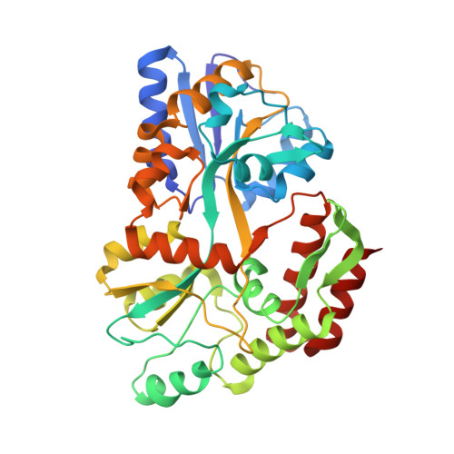
1MDQ

REFINED STRUCTURES OF TWO INSERTION(SLASH)DELETION MUTANTS PROBE FUNCTION OF THE MALTODEXTRIN BINDING PROTEIN


Experimental Data Snapshot

  • Method: X-RAY DIFFRACTION
  • Resolution: 1.90 Å
  • R-Value Work: 0.220 
  • R-Value Observed: 0.220 

wwPDB Validation   3D Report Full Report


This is version 2.1 of the entry. See complete history


Literature

Refined structures of two insertion/deletion mutants probe function of the maltodextrin binding protein.

Sharff, A.J.Rodseth, L.E.Szmelcman, S.Hofnung, M.Quiocho, F.A.

(1995) J Mol Biol 246: 8-13

  • DOI: https://doi.org/10.1006/jmbi.1994.0059
  • Primary Citation of Related Structures:  
    1MDP, 1MDQ

  • PubMed Abstract: 

    The X-ray structures of the maltose bound forms of two insertion/deletion mutants of the Escherichia coli maltodextrin binding protein, MalE322 and MalE178, have been determined and refined. MalE322 involves a one residue deletion, two residue insertion in a hinge segment connecting the two (N and C) domains of the protein, an area already identified as being critical for the correct functioning of the protein. MalE178 involves a nine residue deletion and two residue insertion in a helix at the periphery of the C-domain. The function of both mutant proteins is similar to the wild-type, although MalE322 increases the ability to transport maltose and maltodextrin whilst inhibiting the ability of the cell to grow on dextrins. Both proteins exhibit very localized and conservative conformational changes due to their mutations. The structure of MalE322 shows some deformation of the third hinge strand, indicating the likely cause of change in its biochemistry. MalE178 is stable and its activity virtually unchanged from the wild-type. This is most likely due to the long distance of the mutation from the binding site and conservation of the number of interactions between the area around the deletion site and the main body of the protein.


  • Organizational Affiliation

    Howard Hughes Medical Institute, Baylor College of Medicine, Houston, TX 77030.


Macromolecules
Find similar proteins by:  (by identity cutoff)  |  3D Structure
Entity ID: 1
MoleculeChains Sequence LengthOrganismDetailsImage
MALTODEXTRIN BINDING PROTEIN371Escherichia coliMutation(s): 0 
UniProt
Find proteins for P0AEX9 (Escherichia coli (strain K12))
Explore P0AEX9 
Go to UniProtKB:  P0AEX9
Entity Groups  
Sequence Clusters30% Identity50% Identity70% Identity90% Identity95% Identity100% Identity
UniProt GroupP0AEX9
Sequence Annotations
Expand
  • Reference Sequence
Oligosaccharides

Help

Entity ID: 2
MoleculeChains Length2D Diagram Glycosylation3D Interactions
alpha-D-glucopyranose-(1-4)-alpha-D-glucopyranose
B
2N/A
Glycosylation Resources
GlyTouCan:  G07411ON
GlyCosmos:  G07411ON
Biologically Interesting Molecules (External Reference) 1 Unique
Experimental Data & Validation

Experimental Data

  • Method: X-RAY DIFFRACTION
  • Resolution: 1.90 Å
  • R-Value Work: 0.220 
  • R-Value Observed: 0.220 
  • Space Group: C 1 2 1
Unit Cell:
Length ( Å )Angle ( ˚ )
a = 106.01α = 90
b = 66.46β = 113.17
c = 57.75γ = 90
Software Package:
Software NamePurpose
X-PLORmodel building
PROLSQrefinement
X-PLORrefinement
X-PLORphasing

Structure Validation

View Full Validation Report


Entry History 

Deposition Data

Revision History  (Full details and data files)

  • Version 1.0: 1994-11-01
    Type: Initial release
  • Version 1.1: 2008-03-24
    Changes: Version format compliance
  • Version 1.2: 2011-07-13
    Changes: Version format compliance
  • Version 1.3: 2019-07-17
    Changes: Data collection, Other, Refinement description
  • Version 1.4: 2019-08-14
    Changes: Data collection, Refinement description
  • Version 2.0: 2020-07-29
    Type: Remediation
    Reason: Carbohydrate remediation
    Changes: Advisory, Atomic model, Data collection, Database references, Derived calculations, Non-polymer description, Structure summary
  • Version 2.1: 2024-02-14
    Changes: Data collection, Database references, Structure summary