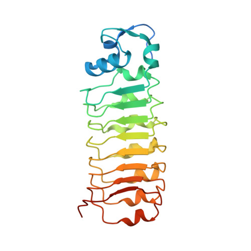
1OTN

Calcium-binding mutant of the Internalin B LRR domain


Experimental Data Snapshot

  • Method: X-RAY DIFFRACTION
  • Resolution: 1.97 Å
  • R-Value Free: 0.244 
  • R-Value Work: 0.199 
  • R-Value Observed: 0.204 

Starting Model: experimental
View more details

wwPDB Validation   3D Report Full Report


This is version 1.5 of the entry. See complete history


Literature

Characterization of the calcium-binding sites of Listeria monocytogenes InlB

Marino, M.Banerjee, M.Copp, J.Dramsi, S.Chapman, T.Van Der Geer, P.Cossart, P.Ghosh, P.

(2004) Biochem Biophys Res Commun 316: 379-386

  • DOI: https://doi.org/10.1016/j.bbrc.2004.02.064
  • Primary Citation of Related Structures:  
    1OTM, 1OTN, 1OTO

  • PubMed Abstract: 

    The Listeria monocytogenes protein InlB promotes invasion of mammalian cells through activation of the receptor tyrosine kinase Met. The InlB N-cap, a approximately 40 residue part of the domain that binds Met, was previously observed to bind two calcium ions in a novel and unusually exposed manner. Because subsequent work raised questions about the existence of these calcium-binding sites, we assayed calcium binding in solution to the InlB N-cap. We show that calcium ions are bound with dissociation constants in the low micromolar range at the two identified sites, and that the sites interact with one another. We demonstrate that the calcium ions are not required for structure, and also find that they have no appreciable effect on Met activation or intracellular invasion. Therefore, our results indicate that the sites are fortuitous in InlB, but also suggest that the simple architecture of the sites may be adaptable for protein engineering purposes.


  • Organizational Affiliation

    Department of Chemistry and Biochemistry, University of California, San Diego, 9500 Gilman Drive, La Jolla, CA 92093-0375, USA.


Macromolecules
Find similar proteins by:  (by identity cutoff)  |  3D Structure
Entity ID: 1
MoleculeChains Sequence LengthOrganismDetailsImage
Internalin B236Listeria monocytogenesMutation(s): 5 
UniProt
Find proteins for P0DQD2 (Listeria monocytogenes serovar 1/2a (strain ATCC BAA-679 / EGD-e))
Explore P0DQD2 
Go to UniProtKB:  P0DQD2
Entity Groups  
Sequence Clusters30% Identity50% Identity70% Identity90% Identity95% Identity100% Identity
UniProt GroupP0DQD2
Sequence Annotations
Expand
  • Reference Sequence
Small Molecules
Ligands 1 Unique
IDChains Name / Formula / InChI Key2D Diagram3D Interactions
CA
Query on CA

Download Ideal Coordinates CCD File 
B [auth A]CALCIUM ION
Ca
BHPQYMZQTOCNFJ-UHFFFAOYSA-N
Experimental Data & Validation

Experimental Data

  • Method: X-RAY DIFFRACTION
  • Resolution: 1.97 Å
  • R-Value Free: 0.244 
  • R-Value Work: 0.199 
  • R-Value Observed: 0.204 
  • Space Group: P 21 21 21
Unit Cell:
Length ( Å )Angle ( ˚ )
a = 44.877α = 90
b = 56.615β = 90
c = 84.03γ = 90
Software Package:
Software NamePurpose
MAR345data collection
SCALEPACKdata scaling
CNSrefinement
CNSphasing

Structure Validation

View Full Validation Report


Entry History 

Deposition Data

Revision History  (Full details and data files)

  • Version 1.0: 2004-03-30
    Type: Initial release
  • Version 1.1: 2008-04-29
    Changes: Version format compliance
  • Version 1.2: 2011-07-13
    Changes: Version format compliance
  • Version 1.3: 2017-10-11
    Changes: Advisory, Refinement description
  • Version 1.4: 2021-10-27
    Changes: Advisory, Database references, Derived calculations
  • Version 1.5: 2023-08-16
    Changes: Data collection, Refinement description