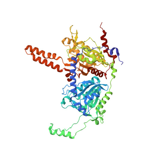
1PIX

Crystal structure of the carboxyltransferase subunit of the bacterial ion pump glutaconyl-coenzyme A decarboxylase


Experimental Data Snapshot

  • Method: X-RAY DIFFRACTION
  • Resolution: 2.20 Å
  • R-Value Free: 0.227 
  • R-Value Work: 0.201 

wwPDB Validation   3D Report Full Report


This is version 1.3 of the entry. See complete history


Literature

Crystal structure of the carboxyltransferase subunit of the bacterial sodium ion pump glutaconyl-coenzyme A decarboxylase

Wendt, K.S.Schall, I.Huber, R.Buckel, W.Jacob, U.

(2003) EMBO J 22: 3493-3502

  • DOI: https://doi.org/10.1093/emboj/cdg358
  • Primary Citation of Related Structures:  
    1PIX

  • PubMed Abstract: 

    Glutaconyl-CoA decarboxylase is a biotin-dependent ion pump whereby the free energy of the glutaconyl-CoA decarboxylation to crotonyl-CoA drives the electrogenic transport of sodium ions from the cytoplasm into the periplasm. Here we present the crystal structure of the decarboxylase subunit (Gcdalpha) from Acidaminococcus fermentans and its complex with glutaconyl-CoA. The active sites of the dimeric Gcdalpha lie at the two interfaces between the mono mers, whereas the N-terminal domain provides the glutaconyl-CoA-binding site and the C-terminal domain binds the biotinyllysine moiety. The Gcdalpha catalyses the transfer of carbon dioxide from glutaconyl-CoA to a biotin carrier (Gcdgamma) that subsequently is decarboxylated by the carboxybiotin decarboxylation site within the actual Na(+) pump (Gcdbeta). The analysis of the active site lead to a novel mechanism for the biotin-dependent carboxy transfer whereby biotin acts as general acid. Furthermore, we propose a holoenzyme assembly in which the water-filled central channel of the Gcdalpha dimer lies co-axial with the ion channel (Gcdbeta). The central channel is blocked by arginines against passage of sodium ions which might enter the central channel through two side channels.


  • Organizational Affiliation

    Max-Planck-Institut für Biochemie, Abteilung Strukturforschung, Am Klopferspitz 18a, D-82152 Martinsried, Germany.


Macromolecules
Find similar proteins by:  (by identity cutoff)  |  3D Structure
Entity ID: 1
MoleculeChains Sequence LengthOrganismDetailsImage
Glutaconyl-CoA decarboxylase A subunit
A, B
587Acidaminococcus fermentansMutation(s): 0 
Gene Names: GCDA
EC: 4.1.1.70 (PDB Primary Data), 7.2.4.5 (UniProt)
UniProt
Find proteins for Q06700 (Acidaminococcus fermentans (strain ATCC 25085 / DSM 20731 / CCUG 9996 / CIP 106432 / VR4))
Explore Q06700 
Go to UniProtKB:  Q06700
Entity Groups  
Sequence Clusters30% Identity50% Identity70% Identity90% Identity95% Identity100% Identity
UniProt GroupQ06700
Sequence Annotations
Expand
  • Reference Sequence
Small Molecules
Ligands 2 Unique
IDChains Name / Formula / InChI Key2D Diagram3D Interactions
SO4
Query on SO4

Download Ideal Coordinates CCD File 
C [auth A],
D [auth A],
H [auth B],
I [auth B],
J [auth B]
SULFATE ION
O4 S
QAOWNCQODCNURD-UHFFFAOYSA-L
FMT
Query on FMT

Download Ideal Coordinates CCD File 
E [auth A],
F [auth A],
G [auth A],
K [auth B]
FORMIC ACID
C H2 O2
BDAGIHXWWSANSR-UHFFFAOYSA-N
Experimental Data & Validation

Experimental Data

  • Method: X-RAY DIFFRACTION
  • Resolution: 2.20 Å
  • R-Value Free: 0.227 
  • R-Value Work: 0.201 
  • Space Group: P 41 21 2
Unit Cell:
Length ( Å )Angle ( ˚ )
a = 151.869α = 90
b = 151.869β = 90
c = 163γ = 90
Software Package:
Software NamePurpose
DENZOdata reduction
SCALEPACKdata scaling
SHARPphasing
CNSrefinement

Structure Validation

View Full Validation Report


Entry History 

Deposition Data

Revision History  (Full details and data files)

  • Version 1.0: 2003-08-05
    Type: Initial release
  • Version 1.1: 2008-04-29
    Changes: Version format compliance
  • Version 1.2: 2011-07-13
    Changes: Version format compliance
  • Version 1.3: 2024-02-14
    Changes: Data collection, Database references, Derived calculations