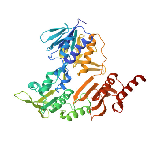
Crystal structure of putidaredoxin reductase from Pseudomonas putida, the final structural component of the cytochrome P450cam monooxygenase.
Sevrioukova, I.F., Li, H., Poulos, T.L.(2004) J Mol Biol 336: 889-902
- PubMed: 15095867
- DOI: https://doi.org/10.1016/j.jmb.2003.12.067
- Primary Citation of Related Structures:
1Q1R, 1Q1W - PubMed Abstract:
The crystal structure of recombinant putidaredoxin reductase (Pdr), an FAD-containing NADH-dependent flavoprotein component of the cytochrome P450cam monooxygenase from Pseudomonas putida, has been determined to 1.90 A resolution. The protein has a fold similar to that of disulfide reductases and consists of the FAD-binding, NAD-binding, and C-terminal domains. Compared to homologous flavoenzymes, the reductase component of biphenyl dioxygenase (BphA4) and apoptosis-inducing factor, Pdr lacks one of the arginine residues that compensates partially for the negative charge on the pyrophosphate of FAD. This uncompensated negative charge is likely to decrease the electron-accepting ability of the flavin. The aromatic side-chain of the "gatekeeper" Tyr159 is in the "out" conformation and leaves the nicotinamide-binding site of Pdr completely open. The presence of electron density in the NAD-binding channel indicates that NAD originating from Escherichia coli is partially bound to Pdr. A structural comparison of Pdr with homologous flavoproteins indicates that an open and accessible nicotinamide-binding site, the presence of an acidic residue in the middle part of the NAD-binding channel that binds the nicotinamide ribose, and multiple positively charged arginine residues surrounding the entrance of the NAD-binding channel are the special structural elements that assist tighter and more specific binding of the oxidized pyridine nucleotide by the BphA4-like flavoproteins. The crystallographic model of Pdr explains differences in the electron transfer mechanism in the Pdr-putidaredoxin redox couple and their mammalian counterparts, adrenodoxin reductase and adrenodoxin.
Organizational Affiliation:
Department of Molecular Biology and Biochemistry, University of California, Irvine, CA 92612-3900, USA. sevrioui@uci.edu


