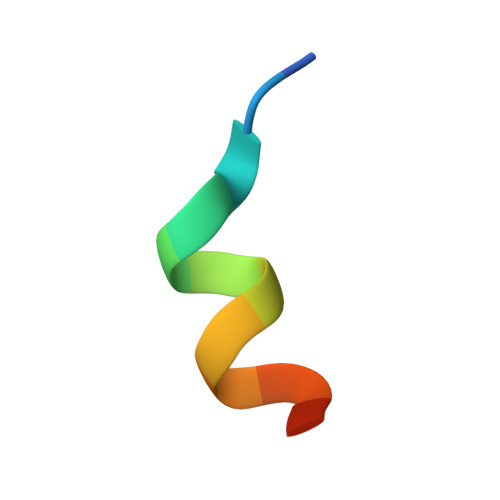
1RKH

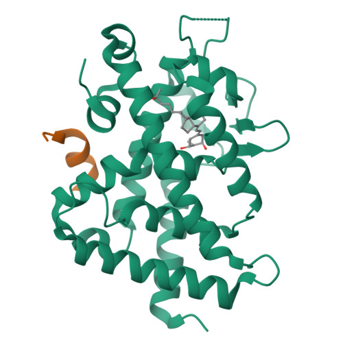
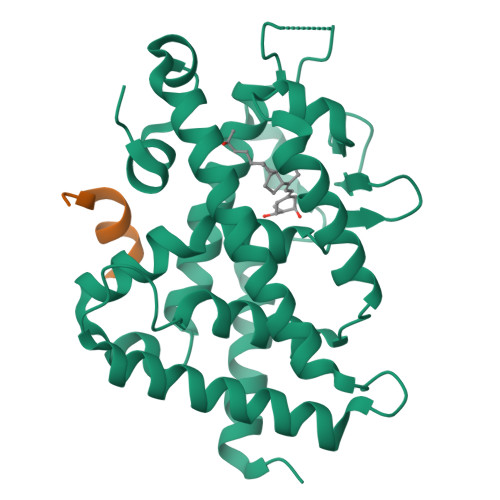
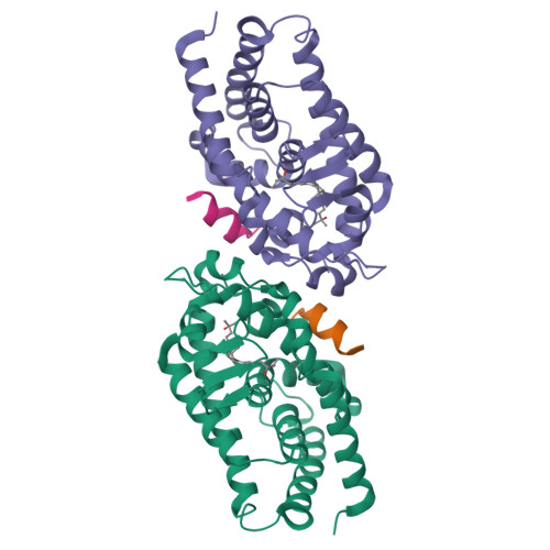
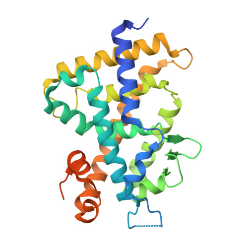
crystal structure of the rat vitamin D receptor ligand binding domain complexed with 2AM20R and a synthetic peptide containing the NR2 box of DRIP 205


Experimental Data Snapshot

  • Method: X-RAY DIFFRACTION
  • Resolution: 2.28 Å
  • R-Value Free: 0.218 
  • R-Value Work: 0.180 
  • R-Value Observed: 0.182 

Starting Model: experimental
View more details

wwPDB Validation   3D Report Full Report


Ligand Structure Quality Assessment 


This is version 1.4 of the entry. See complete history


Literature

Molecular Structure of the Rat Vitamin D Receptor Ligand Binding Domain Complexed with 2-Carbon-Substituted Vitamin D(3) Hormone Analogues and a LXXLL-Containing Coactivator Peptide

Vanhooke, J.L.Benning, M.M.Bauer, C.B.Pike, J.W.DeLuca, H.F.

(2004) Biochemistry 43: 4101-4110

  • DOI: https://doi.org/10.1021/bi036056y
  • Primary Citation of Related Structures:  
    1RJK, 1RK3, 1RKG, 1RKH

  • PubMed Abstract: 

    We have determined the crystal structures of the ligand binding domain (LBD) of the rat vitamin D receptor in ternary complexes with a synthetic LXXLL-containing peptide and the following four ligands: 1alpha,25-dihydroxyvitamin D(3); 2-methylene-19-nor-(20S)-1alpha,25-dihydroxyvitamin D(3) (2MD); 1alpha-hydroxy-2-methylene-19-nor-(20S)-bishomopregnacalciferol (2MbisP), and 2alpha-methyl-19-nor-1alpha,25-dihydroxyvitamin D(3) (2AM20R). The conformation of the LBD is identical in each complex. Binding of the 2-carbon-modified analogues does not change the positions of the amino acids in the ligand binding site and has no effect on the interactions in the coactivator binding pocket. The CD ring of the superpotent analogue, 2MD, is tilted within the binding site relative to the other ligands in this study and to (20S)-1alpha,25-dihydroxyvitamin D(3) [Tocchini-Valentini et al. (2001) Proc. Natl. Acad. Sci. U.S.A. 98, 5491-5496]. The aliphatic side chain of 2MD follows a different path within the binding site; nevertheless, the 25-hydroxyl group at the end of the chain occupies the same position as that of the natural ligand, and the hydrogen bonds with histidines 301 and 393 are maintained. 2MbisP binds to the receptor despite the absence of the 25-hydroxyl group. A water molecule is observed between His 301 and His 393 in this structure, and it preserves the orientation of the histidines in the binding site. Although the alpha-chair conformer is highly favored in solution for the A ring of 2AM20R, the crystal structures demonstrate that this ring assumes the beta-chair conformation in all cases, and the 1alpha-hydroxyl group is equatorial. The peptide folds as a helix and is anchored through hydrogen bonds to a surface groove formed by helices 3, 4, and 12. Electrostatic and hydrophobic interactions between the peptide and the LBD stabilize the active receptor conformation. This stablization appears necessary for crystal growth.


  • Organizational Affiliation

    Department of Biochemistry, University of Wisconsin-Madison, 53711, USA. jlvanhoo@wisc.edu


Macromolecules
Find similar proteins by:  (by identity cutoff)  |  3D Structure
Entity ID: 1
MoleculeChains Sequence LengthOrganismDetailsImage
Vitamin D3 receptor292Rattus norvegicusMutation(s): 0 
Gene Names: VDRNR1I1
UniProt
Find proteins for P13053 (Rattus norvegicus)
Explore P13053 
Go to UniProtKB:  P13053
Entity Groups  
Sequence Clusters30% Identity50% Identity70% Identity90% Identity95% Identity100% Identity
UniProt GroupP13053
Sequence Annotations
Expand
  • Reference Sequence

Find similar proteins by:  Sequence   |   3D Structure  

Entity ID: 2
MoleculeChains Sequence LengthOrganismDetailsImage
Peroxisome proliferator-activated receptor binding proteinB [auth C]13Homo sapiensMutation(s): 0 
Gene Names: MED1
UniProt & NIH Common Fund Data Resources
Find proteins for Q15648 (Homo sapiens)
Explore Q15648 
Go to UniProtKB:  Q15648
PHAROS:  Q15648
GTEx:  ENSG00000125686 
Entity Groups  
Sequence Clusters30% Identity50% Identity70% Identity90% Identity95% Identity100% Identity
UniProt GroupQ15648
Sequence Annotations
Expand
  • Reference Sequence
Small Molecules
Ligands 1 Unique
IDChains Name / Formula / InChI Key2D Diagram3D Interactions
VD2
Query on VD2

Download Ideal Coordinates CCD File 
C [auth A]5-{2-[1-(5-HYDROXY-1,5-DIMETHYL-HEXYL)-7A-METHYL-OCTAHYDRO-INDEN-4-YLIDENE]-ETHYLIDENE}-2-METHYL-CYCLOHEXANE-1,3-DIOL
C27 H46 O3
OZPNRDQPQFOURL-VJPYOZRWSA-N
Experimental Data & Validation

Experimental Data

  • Method: X-RAY DIFFRACTION
  • Resolution: 2.28 Å
  • R-Value Free: 0.218 
  • R-Value Work: 0.180 
  • R-Value Observed: 0.182 
  • Space Group: C 1 2 1
Unit Cell:
Length ( Å )Angle ( ˚ )
a = 154.8α = 90
b = 42.291β = 96.15
c = 41.788γ = 90
Software Package:
Software NamePurpose
REFMACrefinement
SAINTdata reduction
LSCALEdata scaling

Structure Validation

View Full Validation Report


Ligand Structure Quality Assessment 


Entry History 

Deposition Data

Revision History  (Full details and data files)

  • Version 1.0: 2004-04-13
    Type: Initial release
  • Version 1.1: 2008-04-29
    Changes: Version format compliance
  • Version 1.2: 2011-07-13
    Changes: Version format compliance
  • Version 1.3: 2017-10-11
    Changes: Refinement description
  • Version 1.4: 2023-08-23
    Changes: Data collection, Database references, Derived calculations, Refinement description