1S7S

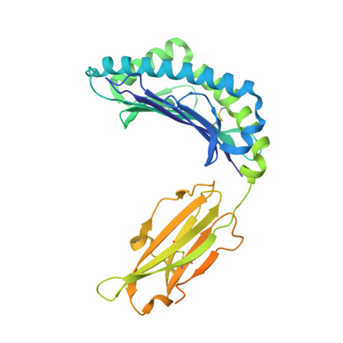
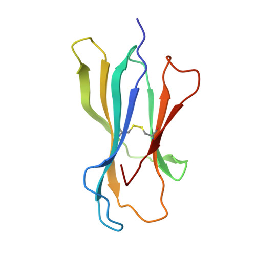
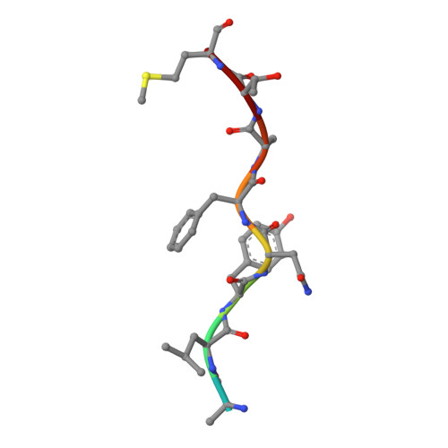
Crystal structures of the murine class I major histocompatibility complex H-2Kb in complex with LCMV-derived gp33 index peptide and three of its escape variants


Experimental Data Snapshot

  • Method: X-RAY DIFFRACTION
  • Resolution: 1.99 Å
  • R-Value Free: 0.232 
  • R-Value Work: 0.190 
  • R-Value Observed: 0.192 

Starting Model: experimental
View more details

wwPDB Validation   3D Report Full Report


This is version 1.5 of the entry. See complete history


Literature

Determination of structural principles underlying three different modes of lymphocytic choriomeningitis virus escape from CTL recognition.

Velloso, L.M.Michaelsson, J.Ljunggren, H.G.Schneider, G.Achour, A.

(2004) J Immunol 172: 5504-5511

  • DOI: https://doi.org/10.4049/jimmunol.172.9.5504
  • Primary Citation of Related Structures:  
    1S7Q, 1S7R, 1S7S, 1S7T, 1S7U, 1S7V, 1S7W, 1S7X

  • PubMed Abstract: 

    Lymphocytic choriomeningitis virus infection of H-2(b) mice generates a strong CD8(+) CTL response mainly directed toward three immunodominant epitopes, one of which, gp33, is presented by both H-2D(b) and H-2K(b) MHC class I molecules. This CTL response acts as a selective agent for the emergence of viral escape variants. These variants generate altered peptide ligands (APLs) that, when presented by class I MHC molecules, antagonize CTL recognition and ultimately allow the virus to evade the cellular immune response. The emergence of APLs of the gp33 epitope is particularly advantageous for LCMV, as it allows viral escape in the context of both H-2D(b) and H-2K(b) MHC class I molecules. We have determined crystal structures of three different APLs of gp33 in complex with both H-2D(b) and H-2K(b). Comparison between these APL/MHC structures and those of the index gp33 peptide/MHC reveals the structural basis for three different strategies used by LCMV viral escape mutations: 1) conformational changes in peptide and MHC residues that are potential TCR contacts, 2) impairment of APL binding to the MHC peptide binding cleft, and 3) introduction of subtle changes at the TCR/pMHC interface, such as the removal of a single hydroxyl group.


  • Organizational Affiliation

    Department of Medical Biochemistry and Biophysics, Karolinska Institute, Stockholm, Sweden.


Macromolecules
Find similar proteins by:  (by identity cutoff)  |  3D Structure
Entity ID: 1
MoleculeChains Sequence LengthOrganismDetailsImage
H-2 class I histocompatibility antigen, K-B alpha chain348Mus musculusMutation(s): 0 
Gene Names: H2-K
UniProt
Find proteins for P01901 (Mus musculus)
Explore P01901 
Go to UniProtKB:  P01901
Entity Groups  
Sequence Clusters30% Identity50% Identity70% Identity90% Identity95% Identity100% Identity
UniProt GroupP01901
Sequence Annotations
Expand
  • Reference Sequence
Find similar proteins by:  (by identity cutoff)  |  3D Structure
Entity ID: 2
MoleculeChains Sequence LengthOrganismDetailsImage
Beta-2-microglobulin99Mus musculusMutation(s): 0 
Gene Names: B2M
UniProt & NIH Common Fund Data Resources
Find proteins for P01887 (Mus musculus)
Explore P01887 
Go to UniProtKB:  P01887
IMPC:  MGI:88127
Entity Groups  
Sequence Clusters30% Identity50% Identity70% Identity90% Identity95% Identity100% Identity
UniProt GroupP01887
Sequence Annotations
Expand
  • Reference Sequence

Find similar proteins by:  Sequence   |   3D Structure  

Entity ID: 3
MoleculeChains Sequence LengthOrganismDetailsImage
Glycoprotein 9-residue peptide9N/AMutation(s): 1 
UniProt
Find proteins for P07399 (Lymphocytic choriomeningitis virus (strain WE))
Explore P07399 
Go to UniProtKB:  P07399
Entity Groups  
UniProt GroupP07399
Sequence Annotations
Expand
  • Reference Sequence
Experimental Data & Validation

Experimental Data

  • Method: X-RAY DIFFRACTION
  • Resolution: 1.99 Å
  • R-Value Free: 0.232 
  • R-Value Work: 0.190 
  • R-Value Observed: 0.192 
  • Space Group: P 21 21 2
Unit Cell:
Length ( Å )Angle ( ˚ )
a = 88.262α = 90
b = 135.555β = 90
c = 45.13γ = 90
Software Package:
Software NamePurpose
REFMACrefinement
HKL-2000data reduction
SCALEPACKdata scaling
AMoREphasing

Structure Validation

View Full Validation Report


Entry History 

Deposition Data

Revision History  (Full details and data files)

  • Version 1.0: 2004-05-04
    Type: Initial release
  • Version 1.1: 2008-04-29
    Changes: Version format compliance
  • Version 1.2: 2011-07-13
    Changes: Advisory, Version format compliance
  • Version 1.3: 2021-10-27
    Changes: Database references
  • Version 1.4: 2023-08-23
    Changes: Data collection, Refinement description
  • Version 1.5: 2024-11-06
    Changes: Structure summary