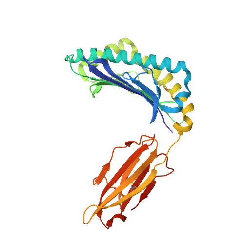
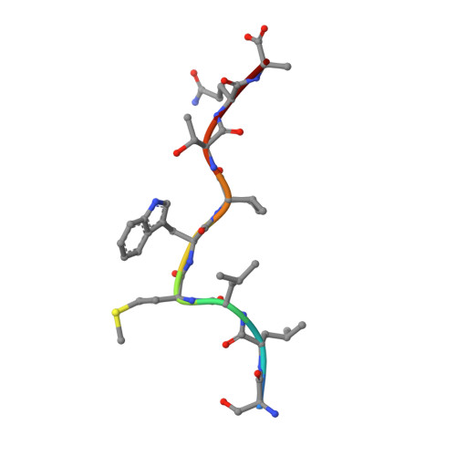
1S9X

Crystal Structure Analysis of NY-ESO-1 epitope analogue, SLLMWITQA, in complex with HLA-A2


Experimental Data Snapshot

  • Method: X-RAY DIFFRACTION
  • Resolution: 2.50 Å
  • R-Value Free: 0.279 
  • R-Value Work: 0.230 

wwPDB Validation   3D Report Full Report


This is version 1.3 of the entry. See complete history


Literature

Functional and structural characteristics of NY-ESO-1-related HLA A2-restricted epitopes and the design of a novel immunogenic analogue

Webb, A.I.Dunstone, M.A.Chen, W.Aguilar, M.I.Chen, Q.Jackson, H.Chang, L.Kjer-Nielsen, L.Beddoe, T.McCluskey, J.Rossjohn, J.Purcell, A.W.

(2004) J Biol Chem 279: 23438-23446

  • DOI: https://doi.org/10.1074/jbc.M314066200
  • Primary Citation of Related Structures:  
    1S9W, 1S9X, 1S9Y

  • PubMed Abstract: 

    NY-ESO-1, a commonly expressed tumor antigen of the cancer-testis family, is expressed by a wide range of tumors but not found in normal adult somatic tissue, making it an ideal cancer vaccine candidate. Peptides derived from NY-ESO-1 have shown preclinical and clinical trial promise; however, biochemical features of these peptides have complicated their formulation and led to heterogeneous immune responses. We have taken a rational approach to engineer an HLA A2-restricted NY-ESO-1-derived T cell epitope with improved formulation and immunogenicity to the wild type peptide. To accomplish this, we have solved the x-ray crystallographic structures of HLA A2 complexed to NY-ESO (157-165) and two analogues of this peptide in which the C-terminal cysteine residue has been substituted to alanine or serine. Substitution of cysteine by serine maintained peptide conformation yet reduced complex stability, resulting in poor cytotoxic T lymphocyte recognition. Conversely, substitution with alanine maintained complex stability and cytotoxic T lymphocyte recognition. Based on the structures of the three HLA A2 complexes, we incorporated 2-aminoisobutyric acid, an isostereomer of cysteine, into the epitope. This analogue is impervious to oxidative damage, cysteinylation, and dimerization of the peptide epitope upon formulation that is characteristic of the wild type peptide. Therefore, this approach has yielded a potential therapeutic molecule that satiates the hydrophobic F pocket of HLA A2 and exhibited superior immunogenicity relative to the wild type peptide.


  • Organizational Affiliation

    Protein Crystallography Unit and Department of Biochemistry and Molecular Biology, School of Biomedical Sciences, Monash University, Clayton, Victoria 3800, Australia.


Macromolecules
Find similar proteins by:  (by identity cutoff)  |  3D Structure
Entity ID: 1
MoleculeChains Sequence LengthOrganismDetailsImage
HLA class I histocompatibility antigen, A-2 alpha chain274Homo sapiensMutation(s): 0 
UniProt & NIH Common Fund Data Resources
Find proteins for P04439 (Homo sapiens)
Explore P04439 
Go to UniProtKB:  P04439
PHAROS:  P04439
GTEx:  ENSG00000206503 
Entity Groups  
Sequence Clusters30% Identity50% Identity70% Identity90% Identity95% Identity100% Identity
UniProt GroupP04439
Sequence Annotations
Expand
  • Reference Sequence
Find similar proteins by:  (by identity cutoff)  |  3D Structure
Entity ID: 2
MoleculeChains Sequence LengthOrganismDetailsImage
Beta-2-microglobulin100Homo sapiensMutation(s): 0 
UniProt & NIH Common Fund Data Resources
Find proteins for P61769 (Homo sapiens)
Explore P61769 
Go to UniProtKB:  P61769
PHAROS:  P61769
GTEx:  ENSG00000166710 
Entity Groups  
Sequence Clusters30% Identity50% Identity70% Identity90% Identity95% Identity100% Identity
UniProt GroupP61769
Sequence Annotations
Expand
  • Reference Sequence

Find similar proteins by:  Sequence   |   3D Structure  

Entity ID: 3
MoleculeChains Sequence LengthOrganismDetailsImage
NY-ESO-1 peptide analogue S9A9N/AMutation(s): 0 
Sequence Annotations
Expand
  • Reference Sequence
Small Molecules
Ligands 1 Unique
IDChains Name / Formula / InChI Key2D Diagram3D Interactions
SO4
Query on SO4

Download Ideal Coordinates CCD File 
D [auth A]SULFATE ION
O4 S
QAOWNCQODCNURD-UHFFFAOYSA-L
Experimental Data & Validation

Experimental Data

  • Method: X-RAY DIFFRACTION
  • Resolution: 2.50 Å
  • R-Value Free: 0.279 
  • R-Value Work: 0.230 
  • Space Group: P 21 3
Unit Cell:
Length ( Å )Angle ( ˚ )
a = 117.95α = 90
b = 117.95β = 90
c = 117.95γ = 90
Software Package:
Software NamePurpose
DENZOdata reduction
SCALEPACKdata scaling
AMoREphasing
CNSrefinement

Structure Validation

View Full Validation Report


Entry History 

Deposition Data

Revision History  (Full details and data files)

  • Version 1.0: 2004-09-28
    Type: Initial release
  • Version 1.1: 2008-04-29
    Changes: Version format compliance
  • Version 1.2: 2011-07-13
    Changes: Version format compliance
  • Version 1.3: 2024-11-06
    Changes: Data collection, Database references, Derived calculations, Structure summary