Understanding protein lids: structural analysis of active hinge mutants in triosephosphate isomerase
Kursula, I., Salin, M., Sun, J., Norledge, B.V., Haapalainen, A.M., Sampson, N.S., Wierenga, R.K.(2004) Protein Eng Des Sel 17: 375-382
- PubMed: 15166315
- DOI: https://doi.org/10.1093/protein/gzh048
- Primary Citation of Related Structures:
1SPQ, 1SQ7, 1SSD, 1SSG, 1SU5, 1SW0, 1SW3, 1SW7 - PubMed Abstract:
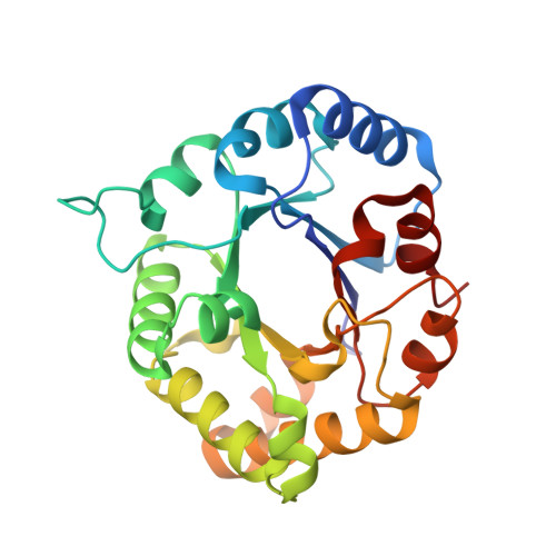
The conformational switch from open to closed of the flexible loop 6 of triosephosphate isomerase (TIM) is essential for the catalytic properties of TIM. Using a directed evolution approach, active variants of chicken TIM with a mutated C-terminal hinge tripeptide of loop 6 have been generated (Sun,J. and Sampson,N.S., Biochemistry, 1999, 38, 11474-11481). In chicken TIM, the wild-type C-terminal hinge tripeptide is KTA. Detailed enzymological characterization of six variants showed that some of these (LWA, NPN, YSL, KTK) have decreased catalytic efficiency, whereas others (KVA, NSS) are essentially identical with wild-type. The structural characterization of these six variants is reported. No significant structural differences compared with the wild-type are found for KVA, NSS and LWA, but substantial structural adaptations are seen for NPN, YSL and KTK. These structural differences can be understood from the buried position of the alanine side chain in the C-hinge position 3 in the open conformation of wild-type loop 6. Replacement of this alanine with a bulky side chain causes the closed conformation to be favored, which correlates with the decreased catalytic efficiency of these variants. The structural context of loop 6 and loop 7 and their sequence conservation in 133 wild-type sequences is also discussed.
Organizational Affiliation:
Department of Biochemistry and Biocenter Oulu, University of Oulu, PO Box 3000, FIN-90014 University of Oulu, Finland.


