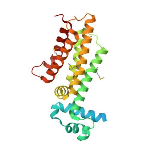
1U9N

Crystal structure of the transcriptional regulator EthR in a ligand bound conformation opens therapeutic perspectives against tuberculosis and leprosy


Experimental Data Snapshot

  • Method: X-RAY DIFFRACTION
  • Resolution: 2.30 Å
  • R-Value Free: 0.231 
  • R-Value Work: 0.201 
  • R-Value Observed: 0.208 

wwPDB Validation   3D Report Full Report


Ligand Structure Quality Assessment 


This is version 1.3 of the entry. See complete history


Literature

Structure of EthR in a Ligand Bound Conformation Reveals Therapeutic Perspectives against Tuberculosis

Frenois, F.Engohang-Ndong, J.Locht, C.Baulard, A.R.Villeret, V.

(2004) Mol Cell 16: 301-307

  • DOI: https://doi.org/10.1016/j.molcel.2004.09.020
  • Primary Citation of Related Structures:  
    1U9N, 1U9O

  • PubMed Abstract: 

    Mycobacterium tuberculosis EthR is a repressor of ethA, a gene encoding a mono-oxygenase required for the activation of the prodrug ethionamide. Here we describe the X-ray crystal structure of EthR, a homodimer with an entirely helical structure showing similarities to TetR family members. Each monomer contained a fortuitous ligand identified as hexadecyl octanoate. The crystal structure of EthR purified in M. smegmatis revealed the presence of a comparable ligand. The binding of hexadecyl octanoate to EthR induces a conformational state incompatible with repressor function, which should lead to ethA derepression and consequently to an increased sensitivity to ethionamide and other thioamides. A related, more hydrophilic ketone was found to exhibit synergistic antimycobacterial effects when tested together with ethionamide, indicating that this strategy may help reduce the dosage of potent antibacterial compounds that otherwise are too toxic to be used as first-line drugs.


  • Organizational Affiliation

    CNRS-UMR8525, 1 rue du Professeur Calmette, BP245 59019 Lille cedex, France.


Macromolecules
Find similar proteins by:  (by identity cutoff)  |  3D Structure
Entity ID: 1
MoleculeChains Sequence LengthOrganismDetailsImage
Transcriptional repressor EthR236Mycobacterium tuberculosisMutation(s): 0 
UniProt
Find proteins for P9WMC1 (Mycobacterium tuberculosis (strain ATCC 25618 / H37Rv))
Explore P9WMC1 
Go to UniProtKB:  P9WMC1
Entity Groups  
Sequence Clusters30% Identity50% Identity70% Identity90% Identity95% Identity100% Identity
UniProt GroupP9WMC1
Sequence Annotations
Expand
  • Reference Sequence
Small Molecules
Ligands 1 Unique
IDChains Name / Formula / InChI Key2D Diagram3D Interactions
CNS
Query on CNS

Download Ideal Coordinates CCD File 
B [auth A]HEXADECYL OCTANOATE
C24 H48 O2
DWMMZQMXUWUJME-UHFFFAOYSA-N
Modified Residues  1 Unique
IDChains TypeFormula2D DiagramParent
MSE
Query on MSE
A
L-PEPTIDE LINKINGC5 H11 N O2 SeMET
Experimental Data & Validation

Experimental Data

  • Method: X-RAY DIFFRACTION
  • Resolution: 2.30 Å
  • R-Value Free: 0.231 
  • R-Value Work: 0.201 
  • R-Value Observed: 0.208 
  • Space Group: P 41 21 2
Unit Cell:
Length ( Å )Angle ( ˚ )
a = 122.38α = 90
b = 122.38β = 90
c = 33.57γ = 90
Software Package:
Software NamePurpose
XDSdata scaling
XDSdata reduction
SHARPphasing
CNSrefinement

Structure Validation

View Full Validation Report


Ligand Structure Quality Assessment 


Entry History 

Deposition Data

Revision History  (Full details and data files)

  • Version 1.0: 2004-11-16
    Type: Initial release
  • Version 1.1: 2008-04-30
    Changes: Version format compliance
  • Version 1.2: 2011-07-13
    Changes: Version format compliance
  • Version 1.3: 2024-11-06
    Changes: Data collection, Database references, Derived calculations, Structure summary